3VQX

Crystal structure of the catalytic domain of pyrrolysyl-tRNA synthetase in triclinic crystal form


Experimental Data Snapshot

  • Method: X-RAY DIFFRACTION
  • Resolution: 2.30 Å
  • R-Value Free: 0.270 
  • R-Value Work: 0.212 
  • R-Value Observed: 0.212 

wwPDB Validation   3D Report Full Report


Ligand Structure Quality Assessment 


This is version 1.2 of the entry. See complete history


Literature

A novel crystal form of pyrrolysyl-tRNA synthetase reveals the pre- and post-aminoacyl-tRNA synthesis conformational states of the adenylate and aminoacyl moieties and an asparagine residue in the catalytic site

Yanagisawa, T.Sumida, T.Ishii, R.Yokoyama, S.

(2013) Acta Crystallogr D Biol Crystallogr 69: 5-15

  • DOI: https://doi.org/10.1107/S0907444912039881
  • Primary Citation of Related Structures:  
    3VQV, 3VQW, 3VQX, 3VQY

  • PubMed Abstract: 

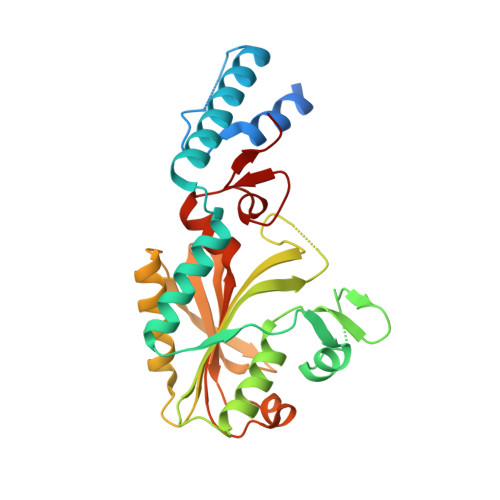
    Structures of Methanosarcina mazei pyrrolysyl-tRNA synthetase (PylRS) have been determined in a novel crystal form. The triclinic form crystals contained two PylRS dimers (four monomer molecules) in the asymmetric unit, in which the two subunits in one dimer each bind N(ℇ)-(tert-butyloxycarbonyl)-L-lysyladenylate (BocLys-AMP) and the two subunits in the other dimer each bind AMP. The BocLys-AMP molecules adopt a curved conformation and the C(α) position of BocLys-AMP protrudes from the active site. The β7-β8 hairpin structures in the four PylRS molecules represent distinct conformations of different states of the aminoacyl-tRNA synthesis reaction. Tyr384, at the tip of the β7-β8 hairpin, moves from the edge to the inside of the active-site pocket and adopts multiple conformations in each state. Furthermore, a new crystal structure of the BocLys-AMPPNP-bound form is also reported. The bound BocLys adopts an unusually bent conformation, which differs from the previously reported structure. It is suggested that the present BocLys-AMPPNP-bound, BocLys-AMP-bound and AMP-bound complexes represent the initial binding of an amino acid (or pre-aminoacyl-AMP synthesis), pre-aminoacyl-tRNA synthesis and post-aminoacyl-tRNA synthesis states, respectively. The conformational changes of Asn346 that accompany the aminoacyl-tRNA synthesis reaction have been captured by X-ray crystallographic analyses. The orientation of the Asn346 side chain, which hydrogen-bonds to the carbonyl group of the amino-acid substrate, shifts by a maximum of 85-90° around the C(β) atom.


  • Organizational Affiliation

    RIKEN Systems and Structural Biology Center, Tsurumi, Yokohama, Japan.


Macromolecules
Find similar proteins by:  (by identity cutoff)  |  3D Structure
Entity ID: 1
MoleculeChains Sequence LengthOrganismDetailsImage
Pyrrolysine--tRNA ligase
A, B, C, D
291Methanosarcina mazeiMutation(s): 0 
Gene Names: pylS
EC: 6.1.1.26
UniProt
Find proteins for Q8PWY1 (Methanosarcina mazei (strain ATCC BAA-159 / DSM 3647 / Goe1 / Go1 / JCM 11833 / OCM 88))
Explore Q8PWY1 
Go to UniProtKB:  Q8PWY1
Entity Groups  
Sequence Clusters30% Identity50% Identity70% Identity90% Identity95% Identity100% Identity
UniProt GroupQ8PWY1
Sequence Annotations
Expand
  • Reference Sequence
Small Molecules
Ligands 3 Unique
IDChains Name / Formula / InChI Key2D Diagram3D Interactions
BLK
Query on BLK

Download Ideal Coordinates CCD File 
I [auth C],
K [auth D]
[[(2R,3S,4R,5R)-5-(6-aminopurin-9-yl)-3,4-bis(oxidanyl)oxolan-2-yl]methoxy-oxidanyl-phosphoryl] (2S)-2-azanyl-6-[(2-methylpropan-2-yl)oxycarbonylamino]hexanoate
C21 H34 N7 O10 P
XYDQQGDGPLGSGU-URQYDQELSA-N
AMP
Query on AMP

Download Ideal Coordinates CCD File 
E [auth A],
G [auth B]
ADENOSINE MONOPHOSPHATE
C10 H14 N5 O7 P
UDMBCSSLTHHNCD-KQYNXXCUSA-N
PO4
Query on PO4

Download Ideal Coordinates CCD File 
F [auth A],
H [auth B],
J [auth C],
L [auth D]
PHOSPHATE ION
O4 P
NBIIXXVUZAFLBC-UHFFFAOYSA-K
Experimental Data & Validation

Experimental Data

  • Method: X-RAY DIFFRACTION
  • Resolution: 2.30 Å
  • R-Value Free: 0.270 
  • R-Value Work: 0.212 
  • R-Value Observed: 0.212 
  • Space Group: P 1
Unit Cell:
Length ( Å )Angle ( ˚ )
a = 62.798α = 106.92
b = 69.422β = 91.41
c = 83.326γ = 113.83
Software Package:
Software NamePurpose
HKL-2000data collection
MOLREPphasing
CNSrefinement
HKL-2000data reduction
HKL-2000data scaling

Structure Validation

View Full Validation Report


Ligand Structure Quality Assessment 


Entry History 

Deposition Data

Revision History  (Full details and data files)

  • Version 1.0: 2013-01-02
    Type: Initial release
  • Version 1.1: 2013-09-04
    Changes: Database references
  • Version 1.2: 2023-11-08
    Changes: Data collection, Database references, Derived calculations, Refinement description