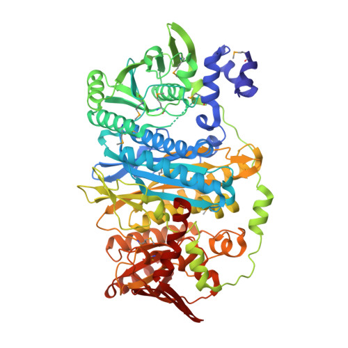
3VIU

Crystal structure of PurL from thermus thermophilus


Experimental Data Snapshot

  • Method: X-RAY DIFFRACTION
  • Resolution: 2.35 Å
  • R-Value Free: 0.260 
  • R-Value Work: 0.199 
  • R-Value Observed: 0.199 

wwPDB Validation   3D Report Full Report


Ligand Structure Quality Assessment 


This is version 1.1 of the entry. See complete history


Literature

Structure of N-formylglycinamide ribonucleotide amidotransferase II (PurL) from Thermus thermophilus HB8

Suzuki, S.Yanai, H.Kanagawa, M.Tamura, S.Watanabe, Y.Fuse, K.Baba, S.Sampei, G.Kawai, G.

(2012) Acta Crystallogr Sect F Struct Biol Cryst Commun 68: 14-19

  • DOI: https://doi.org/10.1107/S1744309111048184
  • Primary Citation of Related Structures:  
    3VIU

  • PubMed Abstract: 

    The crystal structure of PurL from Thermus thermophilus HB8 (TtPurL; TTHA1519) was determined in complex with an adenine nucleotide, PO(4)(3-) and Mg(2+) at 2.35 Å resolution. TtPurL consists of 29 α-helices and 28 β-strands, and one loop is disordered. TtPurL consists of four domains, A1, A2, B1 and B2, and the structures of the A1-B1 and A2-B2 domains were almost identical to each other. Although the sequence identity between TtPurL and PurL from Thermotoga maritima (TmPurL) is higher than that between TtPurL and the PurL domain of the large PurL from Salmonella typhimurium (StPurL), the secondary structure of TtPurL is much more similar to that of StPurL than to that of TmPurL.


  • Organizational Affiliation

    Department of Life and Environmental Sciences, Faculty of Engineering, Chiba Institute of Technology, Narashino, Chiba 275-0016, Japan.


Macromolecules
Find similar proteins by:  (by identity cutoff)  |  3D Structure
Entity ID: 1
MoleculeChains Sequence LengthOrganismDetailsImage
Phosphoribosylformylglycinamidine synthase 2725Thermus thermophilus HB8Mutation(s): 0 
Gene Names: purLTTHA1519
EC: 6.3.5.3
UniProt
Find proteins for Q5SMH8 (Thermus thermophilus (strain ATCC 27634 / DSM 579 / HB8))
Explore Q5SMH8 
Go to UniProtKB:  Q5SMH8
Entity Groups  
Sequence Clusters30% Identity50% Identity70% Identity90% Identity95% Identity100% Identity
UniProt GroupQ5SMH8
Sequence Annotations
Expand
  • Reference Sequence
Small Molecules
Modified Residues  1 Unique
IDChains TypeFormula2D DiagramParent
MSE
Query on MSE
A
L-PEPTIDE LINKINGC5 H11 N O2 SeMET
Experimental Data & Validation

Experimental Data

  • Method: X-RAY DIFFRACTION
  • Resolution: 2.35 Å
  • R-Value Free: 0.260 
  • R-Value Work: 0.199 
  • R-Value Observed: 0.199 
  • Space Group: C 2 2 21
Unit Cell:
Length ( Å )Angle ( ˚ )
a = 94.899α = 90
b = 94.563β = 90
c = 158.78γ = 90
Software Package:
Software NamePurpose
BSSdata collection
SOLVEphasing
CNSrefinement
HKL-2000data reduction
HKL-2000data scaling

Structure Validation

View Full Validation Report


Ligand Structure Quality Assessment 


Entry History 

Deposition Data

Revision History  (Full details and data files)

  • Version 1.0: 2012-01-18
    Type: Initial release
  • Version 1.1: 2012-04-25
    Changes: Database references