3VI8

Human PPAR alpha ligand binding domain in complex with a synthetic agonist APHM13


Experimental Data Snapshot

  • Method: X-RAY DIFFRACTION
  • Resolution: 1.75 Å
  • R-Value Free: 0.249 
  • R-Value Work: 0.214 
  • R-Value Observed: 0.214 

wwPDB Validation   3D Report Full Report


Ligand Structure Quality Assessment 


This is version 1.2 of the entry. See complete history


Literature

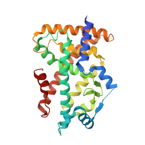
Peroxisome proliferator-activated receptors (PPARs) have multiple binding points that accommodate ligands in various conformations: phenylpropanoic acid-type PPAR ligands bind to PPAR in different conformations, depending on the subtype

Kuwabara, N.Oyama, T.Tomioka, D.Ohashi, M.Yanagisawa, J.Shimizu, T.Miyachi, H.

(2012) J Med Chem 55: 893-902

  • DOI: https://doi.org/10.1021/jm2014293
  • Primary Citation of Related Structures:  
    3VI8, 3VJH, 3VJI

  • PubMed Abstract: 

    Human peroxisome proliferator-activated receptors (hPPARs) are ligand-dependent transcription factors that control various biological responses, and there are three subtypes: hPPARα, hPPARδ, and hPPARγ. We report here that α-substituted phenylpropanoic acid-type hPPAR agonists with similar structure bind to the hPPAR ligand binding domain (LBD) in different conformations, depending on the receptor subtype. These results might indicate that hPPAR ligand binding pockets have multiple binding points that can be utilized to accommodate structurally flexible hPPAR ligands.


  • Organizational Affiliation

    Graduate School of Pharmaceutical Sciences, Faculty of Pharmaceutical Sciences, The University of Tokyo, 7-3-1 Hongo, Bunkyo-ku, Tokyo 113-0033, Japan.


Macromolecules
Find similar proteins by:  (by identity cutoff)  |  3D Structure
Entity ID: 1
MoleculeChains Sequence LengthOrganismDetailsImage
Peroxisome proliferator-activated receptor alpha273Homo sapiensMutation(s): 0 
Gene Names: PPARA
UniProt & NIH Common Fund Data Resources
Find proteins for Q07869 (Homo sapiens)
Explore Q07869 
Go to UniProtKB:  Q07869
PHAROS:  Q07869
GTEx:  ENSG00000186951 
Entity Groups  
Sequence Clusters30% Identity50% Identity70% Identity90% Identity95% Identity100% Identity
UniProt GroupQ07869
Sequence Annotations
Expand
  • Reference Sequence
Small Molecules
Ligands 1 Unique
IDChains Name / Formula / InChI Key2D Diagram3D Interactions
13M
Query on 13M

Download Ideal Coordinates CCD File 
B [auth A](2S)-2-(4-methoxy-3-{[(pyren-1-ylcarbonyl)amino]methyl}benzyl)butanoic acid
C30 H27 N O4
WXYOTFKPGLXJBW-IBGZPJMESA-N
Binding Affinity Annotations 
IDSourceBinding Affinity
13M BindingDB:  3VI8 EC50: 12 (nM) from 1 assay(s)
Experimental Data & Validation

Experimental Data

  • Method: X-RAY DIFFRACTION
  • Resolution: 1.75 Å
  • R-Value Free: 0.249 
  • R-Value Work: 0.214 
  • R-Value Observed: 0.214 
  • Space Group: P 1 21 1
Unit Cell:
Length ( Å )Angle ( ˚ )
a = 44.368α = 90
b = 61.304β = 106.95
c = 53.179γ = 90
Software Package:
Software NamePurpose
HKL-2000data collection
CNSrefinement
HKL-2000data reduction
HKL-2000data scaling
CNSphasing

Structure Validation

View Full Validation Report


Ligand Structure Quality Assessment 


Entry History 

Deposition Data

Revision History  (Full details and data files)

  • Version 1.0: 2012-08-29
    Type: Initial release
  • Version 1.1: 2016-03-02
    Changes: Structure summary
  • Version 1.2: 2023-11-08
    Changes: Data collection, Database references, Derived calculations, Refinement description