


Crystal structure of the Rhodopseudomonas palustris histidine kinase HK9 sensor domain
Zhang, Z., Liu, Q., Hendrickson, W.A.To be published.
Experimental Data Snapshot
wwPDB Validation 3D Report Full Report

Entity ID: 1 | |||||
|---|---|---|---|---|---|
| Molecule | Chains | Sequence Length | Organism | Details | Image |
| Sensor histidine kinase | 164 | Rhodopseudomonas palustris CGA009 | Mutation(s): 0 Gene Names: RPA2292 EC: 2.7.13.3 | 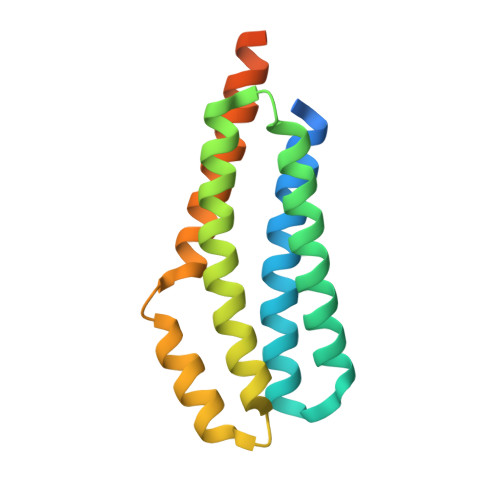 | |
UniProt | |||||
Find proteins for Q6N7G5 (Rhodopseudomonas palustris (strain ATCC BAA-98 / CGA009)) Explore Q6N7G5 Go to UniProtKB: Q6N7G5 | |||||
Entity Groups | |||||
| Sequence Clusters | 30% Identity50% Identity70% Identity90% Identity95% Identity100% Identity | ||||
| UniProt Group | Q6N7G5 | ||||
Sequence AnnotationsExpand | |||||
| |||||
| Ligands 1 Unique | |||||
|---|---|---|---|---|---|
| ID | Chains | Name / Formula / InChI Key | 2D Diagram | 3D Interactions | |
| CL Query on CL | B [auth A] | CHLORIDE ION Cl VEXZGXHMUGYJMC-UHFFFAOYSA-M |  | ||
| Length ( Å ) | Angle ( ˚ ) |
|---|---|
| a = 76.147 | α = 90 |
| b = 76.147 | β = 90 |
| c = 109.155 | γ = 90 |
| Software Name | Purpose |
|---|---|
| ADSC | data collection |
| SHELXD | phasing |
| PHENIX | refinement |
| XDS | data reduction |
| SCALA | data scaling |
RCSB PDB (citation) is hosted by
RCSB PDB is a member of the

