3V7L

Apo Structure of Rat DNA polymerase beta K72E variant


Experimental Data Snapshot

  • Method: X-RAY DIFFRACTION
  • Resolution: 2.66 Å
  • R-Value Free: 0.335 
  • R-Value Work: 0.256 
  • R-Value Observed: 0.260 

wwPDB Validation   3D Report Full Report


This is version 1.2 of the entry. See complete history


Literature

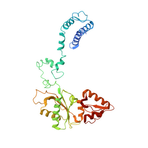
Crystallographic studies of K72E mutant DNA polymerase explain loss of lyase function and reveal changes in the overall conformational state of the polymerase domain

Rangarajan, S.Gridley, C.L.Firbank, S.Dalal, S.Sweasy, J.B.Jaeger, J.

To be published.

Macromolecules
Find similar proteins by:  (by identity cutoff)  |  3D Structure
Entity ID: 1
MoleculeChains Sequence LengthOrganismDetailsImage
DNA polymerase beta340Rattus norvegicusMutation(s): 1 
Gene Names: Polb
EC: 2.7.7.7 (PDB Primary Data), 4.2.99 (PDB Primary Data)
UniProt
Find proteins for P06766 (Rattus norvegicus)
Explore P06766 
Go to UniProtKB:  P06766
Entity Groups  
Sequence Clusters30% Identity50% Identity70% Identity90% Identity95% Identity100% Identity
UniProt GroupP06766
Sequence Annotations
Expand
  • Reference Sequence
Experimental Data & Validation

Experimental Data

  • Method: X-RAY DIFFRACTION
  • Resolution: 2.66 Å
  • R-Value Free: 0.335 
  • R-Value Work: 0.256 
  • R-Value Observed: 0.260 
  • Space Group: P 1 21 1
Unit Cell:
Length ( Å )Angle ( ˚ )
a = 37.425α = 90
b = 71.386β = 90.52
c = 82.905γ = 90
Software Package:
Software NamePurpose
HKL-2000data collection
PHENIXrefinement
HKL-2000data reduction
SCALAdata scaling

Structure Validation

View Full Validation Report


Entry History 

Deposition Data

Revision History  (Full details and data files)

  • Version 1.0: 2013-01-16
    Type: Initial release
  • Version 1.1: 2017-11-08
    Changes: Advisory, Refinement description
  • Version 1.2: 2024-02-28
    Changes: Advisory, Data collection, Database references, Derived calculations