3V6Z

Crystal Structure of Hepatitis B Virus e-antigen


Experimental Data Snapshot

  • Method: X-RAY DIFFRACTION
  • Resolution: 3.34 Å
  • R-Value Free: 0.233 
  • R-Value Work: 0.234 
  • R-Value Observed: 0.234 

wwPDB Validation   3D Report Full Report


This is version 1.1 of the entry. See complete history


Literature

Antigenic switching of hepatitis B virus by alternative dimerization of the capsid protein.

Dimattia, M.A.Watts, N.R.Stahl, S.J.Grimes, J.M.Steven, A.C.Stuart, D.I.Wingfield, P.T.

(2013) Structure 21: 133-142

  • DOI: https://doi.org/10.1016/j.str.2012.10.017
  • Primary Citation of Related Structures:  
    3V6F, 3V6Z

  • PubMed Abstract: 

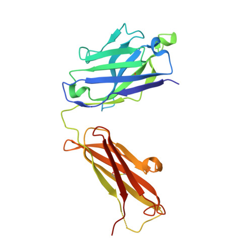
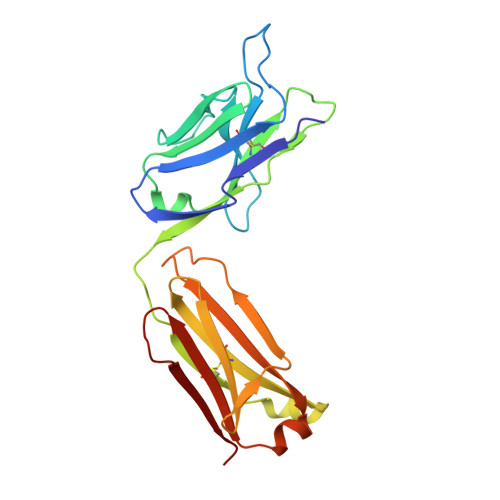
    Chronic hepatitis B virus (HBV) infection afflicts millions worldwide with cirrhosis and liver cancer. HBV e-antigen (HBeAg), a clinical marker for disease severity, is a nonparticulate variant of the protein (core antigen, HBcAg) that forms the building-blocks of capsids. HBeAg is not required for virion production, but is implicated in establishing immune tolerance and chronic infection. Here, we report the crystal structure of HBeAg, which clarifies how the short N-terminal propeptide of HBeAg induces a radically altered mode of dimerization relative to HBcAg (∼140° rotation), locked into place through formation of intramolecular disulfide bridges. This structural switch precludes capsid assembly and engenders a distinct antigenic repertoire, explaining why the two antigens are cross-reactive at the T cell level (through sequence identity) but not at the B cell level (through conformation). The structure offers insight into how HBeAg may establish immune tolerance for HBcAg while evading its robust immunogenicity.


  • Organizational Affiliation

    Division of Structural Biology, Henry Wellcome Building for Genomic Medicine, University of Oxford, Roosevelt Drive, Headington OX3 7BN, UK; Laboratory of Structural Biology Research, National Institute of Arthritis and Musculoskeletal and Skin Diseases; National Institutes of Health, Bethesda, MD 20892, USA.


Macromolecules
Find similar proteins by:  (by identity cutoff)  |  3D Structure
Entity ID: 1
MoleculeChains Sequence LengthOrganismDetailsImage
Fab e6 Heavy Chain
A, C
224Mus musculusMutation(s): 0 
Entity Groups  
Sequence Clusters30% Identity50% Identity70% Identity90% Identity95% Identity100% Identity
Sequence Annotations
Expand
  • Reference Sequence
Find similar proteins by:  (by identity cutoff)  |  3D Structure
Entity ID: 2
MoleculeChains Sequence LengthOrganismDetailsImage
Fab e6 Light Chain
B, D
219Mus musculusMutation(s): 0 
Entity Groups  
Sequence Clusters30% Identity50% Identity70% Identity90% Identity95% Identity100% Identity
Sequence Annotations
Expand
  • Reference Sequence
Find similar proteins by:  (by identity cutoff)  |  3D Structure
Entity ID: 3
MoleculeChains Sequence LengthOrganismDetailsImage
e-antigen
E, F
159Hepatitis B virusMutation(s): 5 
Gene Names: preC/C
UniProt
Find proteins for Q9QMH8 (Hepatitis B virus)
Explore Q9QMH8 
Go to UniProtKB:  Q9QMH8
Entity Groups  
Sequence Clusters30% Identity50% Identity70% Identity90% Identity95% Identity100% Identity
UniProt GroupQ9QMH8
Sequence Annotations
Expand
  • Reference Sequence
Experimental Data & Validation

Experimental Data

  • Method: X-RAY DIFFRACTION
  • Resolution: 3.34 Å
  • R-Value Free: 0.233 
  • R-Value Work: 0.234 
  • R-Value Observed: 0.234 
  • Space Group: P 1
Unit Cell:
Length ( Å )Angle ( ˚ )
a = 66.66α = 96.77
b = 75.76β = 103.81
c = 88.7γ = 116.04
Software Package:
Software NamePurpose
BUSTERrefinement

Structure Validation

View Full Validation Report


Entry History 

Deposition Data

Revision History  (Full details and data files)

  • Version 1.0: 2013-02-06
    Type: Initial release
  • Version 1.1: 2022-12-21
    Changes: Database references