3V50

Complex of SHV S130G mutant beta-lactamase complexed to SA2-13


Experimental Data Snapshot

  • Method: X-RAY DIFFRACTION
  • Resolution: 1.45 Å
  • R-Value Free: 0.197 
  • R-Value Work: 0.180 
  • R-Value Observed: 0.181 

wwPDB Validation   3D Report Full Report


Ligand Structure Quality Assessment 


This is version 1.0 of the entry. See complete history


Literature

The importance of the trans-enamine intermediate as a beta-lactamase inhibition strategy probed in inhibitor-resistant SHV beta-lactamase variants.

Ke, W.Rodkey, E.A.Sampson, J.M.Skalweit, M.J.Sheri, A.Pagadala, S.R.Nottingham, M.D.Buynak, J.D.Bonomo, R.A.van den Akker, F.

(2012) ChemMedChem 7: 1002-1008

  • DOI: https://doi.org/10.1002/cmdc.201200006
  • Primary Citation of Related Structures:  
    3V50, 3V5M

  • PubMed Abstract: 

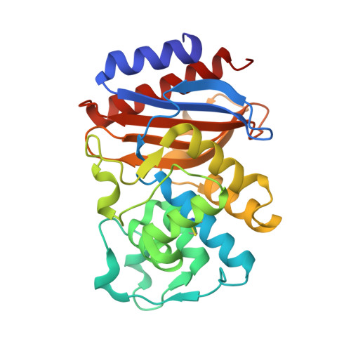
    The ability of bacteria to express inhibitor-resistant (IR) β-lactamases is stimulating the development of novel inhibitors of these enzymes. The 2'β-glutaroxypenicillinate sulfone, SA2-13, was previously designed to enhance the stabilization of the deacylation-refractory, trans-enamine inhibitory intermediate. To test whether this mode of inhibition can overcome different IR mutations, we determined the binding mode of SA2-13 through X-ray crystallography, obtaining co-crystals of the inhibitor-protein complex by soaking crystals of the IR sulfhydryl variable (SHV) β-lactamase variants S130G and M69V with the inhibitor. The 1.45 Å crystal structure of the S130G SHV:SA2-13 complex reveals that SA2-13 is still able to form the stable trans-enamine intermediate similar to the wild-type complex structure, yet with its carboxyl linker shifted deeper into the active site in the space vacated by the S130G mutation. In contrast, data from crystals of the M69V SHV:SA2-13 complex at 1.3 Å did not reveal clear inhibitor density indicating that this IR variant disfavors the trans-enamine conformation, likely due to a subtle shift in A237.


  • Organizational Affiliation

    Department of Biochemistry, RT500, School of Medicine, Case Western Reserve University, 10900 Euclid Avenue, Cleveland, OH 44106, USA.


Macromolecules
Find similar proteins by:  (by identity cutoff)  |  3D Structure
Entity ID: 1
MoleculeChains Sequence LengthOrganismDetailsImage
Beta-lactamase265Klebsiella pneumoniaeMutation(s): 1 
Gene Names: blaSHV-1blaCTX-MblaSHVblaSHV-11blaSHV1SHVshv-1SHV-1b-a
EC: 3.5.2.6
UniProt
Find proteins for P0AD64 (Klebsiella pneumoniae)
Explore P0AD64 
Go to UniProtKB:  P0AD64
Entity Groups  
Sequence Clusters30% Identity50% Identity70% Identity90% Identity95% Identity100% Identity
UniProt GroupP0AD64
Sequence Annotations
Expand
  • Reference Sequence
Small Molecules
Ligands 2 Unique
IDChains Name / Formula / InChI Key2D Diagram3D Interactions
MA4
Query on MA4

Download Ideal Coordinates CCD File 
C [auth A],
D [auth A]
CYCLOHEXYL-HEXYL-BETA-D-MALTOSIDE
C24 H44 O11
WUCWJXGMSXTDAV-QKMCSOCLSA-N
SA2
Query on SA2

Download Ideal Coordinates CCD File 
B [auth A](3R)-4-[(4-CARBOXYBUTANOYL)OXY]-N-[(1E)-3-OXOPROP-1-EN-1-YL]-3-SULFINO-D-VALINE
C13 H19 N O9 S
ZLJAMSCQNNEARN-QDZHIHTESA-N
Experimental Data & Validation

Experimental Data

  • Method: X-RAY DIFFRACTION
  • Resolution: 1.45 Å
  • R-Value Free: 0.197 
  • R-Value Work: 0.180 
  • R-Value Observed: 0.181 
  • Space Group: P 21 21 21
Unit Cell:
Length ( Å )Angle ( ˚ )
a = 49.671α = 90
b = 55.479β = 90
c = 83.62γ = 90
Software Package:
Software NamePurpose
d*TREKdata scaling
PHASERphasing
REFMACrefinement
d*TREKdata reduction

Structure Validation

View Full Validation Report


Ligand Structure Quality Assessment 


Entry History 

Deposition Data

Revision History  (Full details and data files)

  • Version 1.0: 2012-08-08
    Type: Initial release