An amino acid position at crossroads of evolution of protein function: antibiotic sensor domain of BlaR1 protein from Staphylococcus aureus versus clasS D beta-lactamases
Kumarasiri, M., Llarrull, L.I., Borbulevych, O., Fishovitz, J., Lastochkin, E., Baker, B.M., Mobashery, S.(2012) J Biol Chem 287: 8232-8241
- PubMed: 22262858
- DOI: https://doi.org/10.1074/jbc.M111.333179
- Primary Citation of Related Structures:
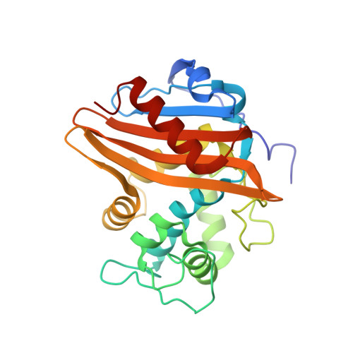
3UY6 - PubMed Abstract:
The integral membrane protein BlaR1 of Staphylococcus aureus senses the presence of β-lactam antibiotics in the milieu and transduces the information to its cytoplasmic side, where its activity unleashes the expression of a set of genes, including that for BlaR1 itself, which manifest the antibiotic-resistant phenotype. The x-ray structure of the sensor domain of this protein exhibits an uncanny similarity to those of the class D β-lactamases. The former is a membrane-bound receptor/sensor for the β-lactam antibiotics, devoid of catalytic competence for substrate turnover, whereas the latter are soluble periplasmic enzymes in gram-negative bacteria with avid ability for β-lactam turnover. The two are clearly related to each other from an evolutionary point of view. However, the high resolution x-ray structures for both by themselves do not reveal why one is a receptor and the other an enzyme. It is documented herein that a single amino acid change at position 439 of the BlaR1 protein is sufficient to endow the receptor/sensor protein with modest turnover ability for cephalosporins as substrates. The x-ray structure for this mutant protein and the dynamics simulations revealed how a hydrolytic water molecule may sequester itself in the antibiotic-binding site to enable hydrolysis of the acylated species. These studies document how the nature of the residue at position 439 is critical for the fate of the protein in imparting unique functions on the same molecular template, to result in one as a receptor and in another as a catalyst.
Organizational Affiliation:
Department of Chemistry and Biochemistry, University of Notre Dame, Notre Dame, Indiana 46556, USA.