3UQ8

Structure of adenylation domain of Haemophilus influenzae DNA ligases bound to NAD+ in adenylated state.


Experimental Data Snapshot

  • Method: X-RAY DIFFRACTION
  • Resolution: 1.70 Å
  • R-Value Free: 0.216 
  • R-Value Work: 0.185 
  • R-Value Observed: 0.187 

wwPDB Validation   3D Report Full Report


Ligand Structure Quality Assessment 


This is version 1.2 of the entry. See complete history


Literature

Structure Guided Understanding of NAD(+) Recognition in Bacterial DNA Ligases.

Lahiri, S.D.Gu, R.F.Gao, N.Karantzeni, I.Walkup, G.K.Mills, S.D.

(2012) ACS Chem Biol 7: 571-580

  • DOI: https://doi.org/10.1021/cb200392g
  • Primary Citation of Related Structures:  
    3UQ8

  • PubMed Abstract: 

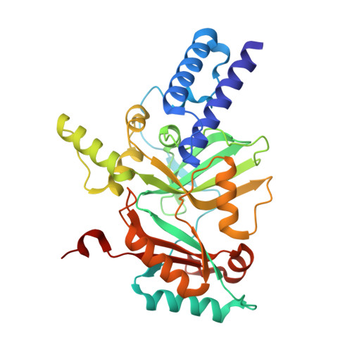
    NAD(+)-dependent DNA ligases (LigA) are essential bacterial enzymes that catalyze phosphodiester bond formation during DNA replication and repair processes. Phosphodiester bond formation proceeds through a 3-step reaction mechanism. In the first step, the LigA adenylation domain interacts with NAD(+) to form a covalent enzyme-AMP complex. Although it is well established that the specificity for binding of NAD(+) resides within the adenylation domain, the precise recognition elements for the initial binding event remain unclear. We report here the structure of the adenylation domain from Haemophilus influenzae LigA. This structure is a first snapshot of a LigA-AMP intermediate with NAD(+) bound to domain 1a in its open conformation. The binding affinities of NAD(+) for adenylated and nonadenylated forms of the H. influenzae LigA adenylation domain were similar. The combined crystallographic and NAD(+)-binding data suggest that the initial recognition of NAD(+) is via the NMN binding region in domain 1a of LigA.


  • Organizational Affiliation

    Department of Bioscience, Infection Innovative Medicines Unit, AstraZeneca R&D Boston, Waltham, Massachusetts 02451, United States. sushmita.lahiri@astrazeneca.com


Macromolecules
Find similar proteins by:  (by identity cutoff)  |  3D Structure
Entity ID: 1
MoleculeChains Sequence LengthOrganismDetailsImage
DNA ligase322Haemophilus influenzae Rd KW20Mutation(s): 0 
Gene Names: ligAligligNHI_1100
EC: 6.5.1.2
UniProt
Find proteins for P43813 (Haemophilus influenzae (strain ATCC 51907 / DSM 11121 / KW20 / Rd))
Explore P43813 
Go to UniProtKB:  P43813
Entity Groups  
Sequence Clusters30% Identity50% Identity70% Identity90% Identity95% Identity100% Identity
UniProt GroupP43813
Sequence Annotations
Expand
  • Reference Sequence
Small Molecules
Ligands 2 Unique
IDChains Name / Formula / InChI Key2D Diagram3D Interactions
NAD
Query on NAD

Download Ideal Coordinates CCD File 
B [auth A]NICOTINAMIDE-ADENINE-DINUCLEOTIDE
C21 H27 N7 O14 P2
BAWFJGJZGIEFAR-NNYOXOHSSA-N
AMP
Query on AMP

Download Ideal Coordinates CCD File 
C [auth A]ADENOSINE MONOPHOSPHATE
C10 H14 N5 O7 P
UDMBCSSLTHHNCD-KQYNXXCUSA-N
Binding Affinity Annotations 
IDSourceBinding Affinity
NAD Binding MOAD:  3UQ8 Kd: 4.50e+4 (nM) from 1 assay(s)
Experimental Data & Validation

Experimental Data

  • Method: X-RAY DIFFRACTION
  • Resolution: 1.70 Å
  • R-Value Free: 0.216 
  • R-Value Work: 0.185 
  • R-Value Observed: 0.187 
  • Space Group: P 43 21 2
Unit Cell:
Length ( Å )Angle ( ˚ )
a = 70.233α = 90
b = 70.233β = 90
c = 161.286γ = 90
Software Package:
Software NamePurpose
MOSFLMdata reduction
REFMACrefinement
PDB_EXTRACTdata extraction
HKL-2000data collection
SCALAdata scaling
AMoREphasing

Structure Validation

View Full Validation Report


Ligand Structure Quality Assessment 


Entry History 

Deposition Data

  • Released Date: 2012-01-25 
  • Deposition Author(s): Lahiri, S.D.

Revision History  (Full details and data files)

  • Version 1.0: 2012-01-25
    Type: Initial release
  • Version 1.1: 2012-03-28
    Changes: Database references
  • Version 1.2: 2014-11-12
    Changes: Structure summary