3UNK

CDK2 in complex with inhibitor YL5-083


Experimental Data Snapshot

  • Method: X-RAY DIFFRACTION
  • Resolution: 2.10 Å
  • R-Value Free: 0.234 
  • R-Value Work: 0.180 

wwPDB Validation   3D Report Full Report


This is version 1.2 of the entry. See complete history


Literature

A Novel Mechanism by Which Small Molecule Inhibitors Induce the DFG Flip in Aurora A.

Martin, M.P.Zhu, J.Y.Lawrence, H.R.Pireddu, R.Luo, Y.Alam, R.Ozcan, S.Sebti, S.M.Lawrence, N.J.Schonbrunn, E.

(2012) ACS Chem Biol 7: 698-706

  • DOI: https://doi.org/10.1021/cb200508b
  • Primary Citation of Related Structures:  
    3UNJ, 3UNK, 3UNZ, 3UO4, 3UO5, 3UO6, 3UOD, 3UOH, 3UOJ, 3UOK, 3UOL, 3UP2

  • PubMed Abstract: 

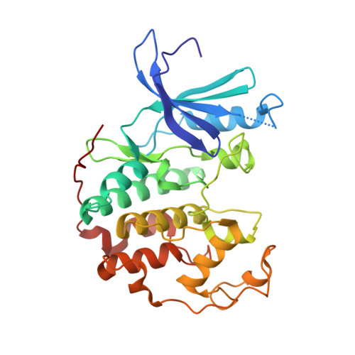
    Most protein kinases share a DFG (Asp-Phe-Gly) motif in the ATP site that can assume two distinct conformations, the active DFG-in and the inactive DFG-out states. Small molecule inhibitors able to induce the DFG-out state have received considerable attention in kinase drug discovery. Using a typical DFG-in inhibitor scaffold of Aurora A, a kinase involved in the regulation of cell division, we found that halogen and nitrile substituents directed at the N-terminally flanking residue Ala273 induced global conformational changes in the enzyme, leading to DFG-out inhibitors that are among the most potent Aurora A inhibitors reported to date. The data suggest an unprecedented mechanism of action, in which induced-dipole forces along the Ala273 side chain alter the charge distribution of the DFG backbone, allowing the DFG to unwind. As the ADFG sequence and three-dimensional structure is highly conserved, DFG-out inhibitors of other kinases may be designed by specifically targeting the flanking alanine residue with electric dipoles.


  • Organizational Affiliation

    Drug Discovery Department, Moffitt Cancer Center and Research Institute, Tampa, Florida 33612, United States.


Macromolecules
Find similar proteins by:  (by identity cutoff)  |  3D Structure
Entity ID: 1
MoleculeChains Sequence LengthOrganismDetailsImage
Cyclin-dependent kinase 2298Homo sapiensMutation(s): 0 
Gene Names: CDK2
EC: 2.7.11.22
UniProt & NIH Common Fund Data Resources
Find proteins for P24941 (Homo sapiens)
Explore P24941 
Go to UniProtKB:  P24941
PHAROS:  P24941
GTEx:  ENSG00000123374 
Entity Groups  
Sequence Clusters30% Identity50% Identity70% Identity90% Identity95% Identity100% Identity
UniProt GroupP24941
Sequence Annotations
Expand
  • Reference Sequence
Small Molecules
Ligands 2 Unique
IDChains Name / Formula / InChI Key2D Diagram3D Interactions
0BY
Query on 0BY

Download Ideal Coordinates CCD File 
B [auth A]4-({4-[(2-chlorophenyl)amino]pyrimidin-2-yl}amino)benzoic acid
C17 H13 Cl N4 O2
BAZFHAGOCKJYHX-UHFFFAOYSA-N
PO4
Query on PO4

Download Ideal Coordinates CCD File 
C [auth A]PHOSPHATE ION
O4 P
NBIIXXVUZAFLBC-UHFFFAOYSA-K
Binding Affinity Annotations 
IDSourceBinding Affinity
0BY Binding MOAD:  3UNK IC50: 1.50e+4 (nM) from 1 assay(s)
PDBBind:  3UNK IC50: 1.50e+4 (nM) from 1 assay(s)
Experimental Data & Validation

Experimental Data

  • Method: X-RAY DIFFRACTION
  • Resolution: 2.10 Å
  • R-Value Free: 0.234 
  • R-Value Work: 0.180 
  • Space Group: P 21 21 21
Unit Cell:
Length ( Å )Angle ( ˚ )
a = 52.95α = 90
b = 72.02β = 90
c = 72.37γ = 90
Software Package:
Software NamePurpose
StructureStudiodata collection
PHENIXmodel building
PHENIXrefinement
XDSdata reduction
XDSdata scaling
PHENIXphasing

Structure Validation

View Full Validation Report


Entry History 

Deposition Data

Revision History  (Full details and data files)

  • Version 1.0: 2012-01-25
    Type: Initial release
  • Version 1.1: 2012-05-02
    Changes: Database references
  • Version 1.2: 2023-09-13
    Changes: Data collection, Database references, Derived calculations, Refinement description