3ULP

Plasmodium falciparum SSB complex with ssDNA


Experimental Data Snapshot

  • Method: X-RAY DIFFRACTION
  • Resolution: 2.10 Å
  • R-Value Free: 0.276 
  • R-Value Work: 0.218 
  • R-Value Observed: 0.221 

wwPDB Validation   3D Report Full Report


This is version 1.2 of the entry. See complete history


Literature

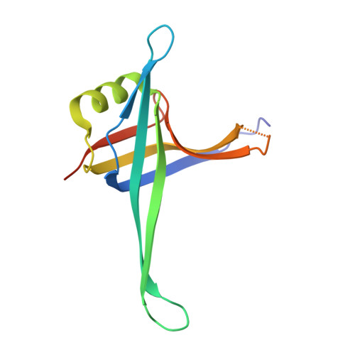
Plasmodium falciparum SSB Tetramer Wraps Single-Stranded DNA with Similar Topology but Opposite Polarity to E. coli SSB.

Antony, E.Weiland, E.A.Korolev, S.Lohman, T.M.

(2012) J Mol Biol 420: 269-283

  • DOI: https://doi.org/10.1016/j.jmb.2012.04.021
  • Primary Citation of Related Structures:  
    3ULP

  • PubMed Abstract: 

    Single-stranded DNA binding (SSB) proteins play central roles in genome maintenance in all organisms. Plasmodium falciparum, the causative agent of malaria, encodes an SSB protein that localizes to the apicoplast and likely functions in the replication and maintenance of its genome. P. falciparum SSB (Pf-SSB) shares a high degree of sequence homology with bacterial SSB proteins but differs in the composition of its C-terminus, which interacts with more than a dozen other proteins in Escherichia coli SSB (Ec-SSB). Using sedimentation methods, we show that Pf-SSB forms a stable homo-tetramer alone and when bound to single-stranded DNA (ssDNA). We also present a crystal structure at 2.1 Å resolution of the Pf-SSB tetramer bound to two (dT)(35) molecules. The Pf-SSB tetramer is structurally similar to the Ec-SSB tetramer, and ssDNA wraps completely around the tetramer with a "baseball seam" topology that is similar to Ec-SSB in its "65 binding mode". However, the polarity of the ssDNA wrapping around Pf-SSB is opposite to that observed for Ec-SSB. The interactions between the bases in the DNA and the amino acid side chains also differ from those observed in the Ec-SSB-DNA structure, suggesting that other differences may exist in the DNA binding properties of these structurally similar proteins.


  • Organizational Affiliation

    Department of Biochemistry and Molecular Biophysics, Washington University School of Medicine, 660 South Euclid Avenue, Box 8231, St. Louis, MO 63110-1093, USA.


Macromolecules

Find similar proteins by:  (by identity cutoff)  |  3D Structure
Entity ID: 1
MoleculeChains Sequence LengthOrganismDetailsImage
Single-strand binding protein
A, B, C, D
124Plasmodium falciparumMutation(s): 0 
Gene Names: PFE0435c
UniProt
Find proteins for Q8I415 (Plasmodium falciparum (isolate 3D7))
Explore Q8I415 
Go to UniProtKB:  Q8I415
Entity Groups  
Sequence Clusters30% Identity50% Identity70% Identity90% Identity95% Identity100% Identity
UniProt GroupQ8I415
Sequence Annotations
Expand
  • Reference Sequence
Find similar nucleic acids by:  (by identity cutoff)  |  3D Structure
Entity ID: 2
MoleculeChains LengthOrganismImage
DNA (35-MER)E [auth R],
F [auth Q]
35synthetic construct
Sequence Annotations
Expand
  • Reference Sequence
Experimental Data & Validation

Experimental Data

  • Method: X-RAY DIFFRACTION
  • Resolution: 2.10 Å
  • R-Value Free: 0.276 
  • R-Value Work: 0.218 
  • R-Value Observed: 0.221 
  • Space Group: C 1 2 1
Unit Cell:
Length ( Å )Angle ( ˚ )
a = 118.017α = 90
b = 82.794β = 99.57
c = 87.556γ = 90
Software Package:
Software NamePurpose
CrystalCleardata collection
PHASESphasing
ARP/wARPmodel building
Cootmodel building
REFMACrefinement
HKL-2000data reduction
HKL-2000data scaling

Structure Validation

View Full Validation Report


Entry History 

Deposition Data

Revision History  (Full details and data files)

  • Version 1.0: 2012-05-09
    Type: Initial release
  • Version 1.1: 2012-07-25
    Changes: Database references
  • Version 1.2: 2023-09-13
    Changes: Data collection, Database references, Refinement description