3UGC

Structural basis of Jak2 inhibition by the type II inhibtor NVP-BBT594


Experimental Data Snapshot

  • Method: X-RAY DIFFRACTION
  • Resolution: 1.34 Å
  • R-Value Free: 0.189 
  • R-Value Work: 0.176 
  • R-Value Observed: 0.177 

wwPDB Validation   3D Report Full Report


Ligand Structure Quality Assessment 


This is version 1.4 of the entry. See complete history


Literature

Modulation of activation-loop phosphorylation by JAK inhibitors is binding mode dependent.

Andraos, R.Qian, Z.Bonenfant, D.Rubert, J.Vangrevelinghe, E.Scheufler, C.Marque, F.Regnier, C.H.De Pover, A.Ryckelynck, H.Bhagwat, N.Koppikar, P.Goel, A.Wyder, L.Tavares, G.Baffert, F.Pissot-Soldermann, C.Manley, P.W.Gaul, C.Voshol, H.Levine, R.L.Sellers, W.R.Hofmann, F.Radimerski, T.

(2012) Cancer Discov 2: 512-523

  • DOI: https://doi.org/10.1158/2159-8290.CD-11-0324
  • Primary Citation of Related Structures:  
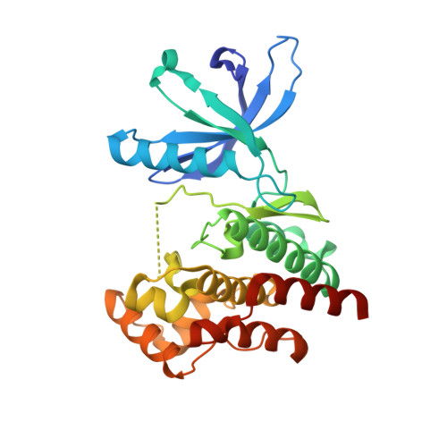
    3UGC

  • PubMed Abstract: 

    Janus kinase (JAK) inhibitors are being developed for the treatment of rheumatoid arthritis, psoriasis, myeloproliferative neoplasms, and leukemias. Most of these drugs target the ATP-binding pocket and stabilize the active conformation of the JAK kinases. This type I binding mode can lead to an increase in JAK activation loop phosphorylation, despite blockade of kinase function. Here we report that stabilizing the inactive state via type II inhibition acts in the opposite manner, leading to a loss of activation loop phosphorylation. We used X-ray crystallography to corroborate the binding mode and report for the first time the crystal structure of the JAK2 kinase domain in an inactive conformation. Importantly, JAK inhibitor-induced activation loop phosphorylation requires receptor interaction, as well as intact kinase and pseudokinase domains. Hence, depending on the respective conformation stabilized by a JAK inhibitor, hyperphosphorylation of the activation loop may or may not be elicited.


  • Organizational Affiliation

    Disease Area Oncology, Novartis Institutes for BioMedical Research, Basel, Switzerland.


Macromolecules
Find similar proteins by:  (by identity cutoff)  |  3D Structure
Entity ID: 1
MoleculeChains Sequence LengthOrganismDetailsImage
Tyrosine-protein kinase JAK2295Homo sapiensMutation(s): 2 
EC: 2.7.10.2
UniProt & NIH Common Fund Data Resources
Find proteins for O60674 (Homo sapiens)
Explore O60674 
Go to UniProtKB:  O60674
PHAROS:  O60674
GTEx:  ENSG00000096968 
Entity Groups  
Sequence Clusters30% Identity50% Identity70% Identity90% Identity95% Identity100% Identity
UniProt GroupO60674
Sequence Annotations
Expand
  • Reference Sequence
Small Molecules
Ligands 2 Unique
IDChains Name / Formula / InChI Key2D Diagram3D Interactions
046
Query on 046

Download Ideal Coordinates CCD File 
B [auth A]5-{[6-(acetylamino)pyrimidin-4-yl]oxy}-N-{4-[(4-methylpiperazin-1-yl)methyl]-3-(trifluoromethyl)phenyl}-2,3-dihydro-1H-indole-1-carboxamide
C28 H30 F3 N7 O3
VQLNKQZLPGLOSI-UHFFFAOYSA-N
MLI
Query on MLI

Download Ideal Coordinates CCD File 
C [auth A]MALONATE ION
C3 H2 O4
OFOBLEOULBTSOW-UHFFFAOYSA-L
Experimental Data & Validation

Experimental Data

  • Method: X-RAY DIFFRACTION
  • Resolution: 1.34 Å
  • R-Value Free: 0.189 
  • R-Value Work: 0.176 
  • R-Value Observed: 0.177 
  • Space Group: C 1 2 1
Unit Cell:
Length ( Å )Angle ( ˚ )
a = 106.97α = 90
b = 69.39β = 98.98
c = 50.8γ = 90
Software Package:
Software NamePurpose
PHASERphasing
BUSTERrefinement
XDSdata reduction
XSCALEdata scaling

Structure Validation

View Full Validation Report


Ligand Structure Quality Assessment 


Entry History 

Deposition Data

Revision History  (Full details and data files)

  • Version 1.0: 2012-05-16
    Type: Initial release
  • Version 1.1: 2012-07-04
    Changes: Non-polymer description
  • Version 1.2: 2012-08-29
    Changes: Database references
  • Version 1.3: 2018-04-11
    Changes: Data collection, Structure summary
  • Version 1.4: 2023-09-13
    Changes: Data collection, Database references, Derived calculations, Refinement description