3UBX

Crystal structure of the mouse CD1d-C20:2-aGalCer-L363 mAb Fab complex


Experimental Data Snapshot

  • Method: X-RAY DIFFRACTION
  • Resolution: 3.10 Å
  • R-Value Free: 0.230 
  • R-Value Work: 0.202 
  • R-Value Observed: 0.203 

wwPDB Validation   3D Report Full Report


Ligand Structure Quality Assessment 


This is version 2.2 of the entry. See complete history


Literature

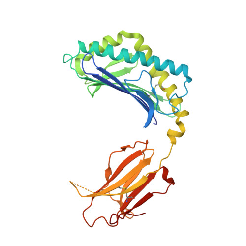
Structural basis for the recognition of C20:2-alpha GalCer by the invariant natural killer T cell receptor-like antibody L363.

Yu, E.D.Girardi, E.Wang, J.Mac, T.T.Yu, K.O.Van Calenbergh, S.Porcelli, S.A.Zajonc, D.M.

(2012) J Biol Chem 287: 1269-1278

  • DOI: https://doi.org/10.1074/jbc.M111.308783
  • Primary Citation of Related Structures:  
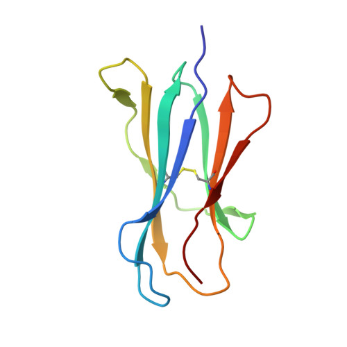
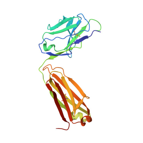
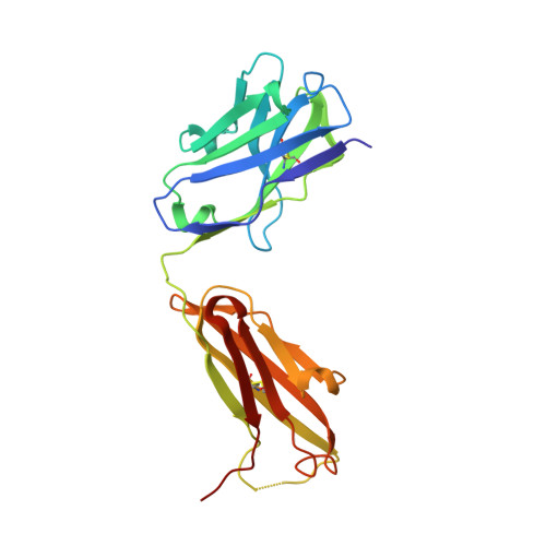
    3UBX

  • PubMed Abstract: 

    Natural killer T (NKT) cells express a semi-invariant Vα14 T cell receptor (TCR) and recognize structurally diverse antigens presented by the antigen-presenting molecule CD1d that range from phosphoglycerolipids to α- and β-anomeric glycosphingolipids, as well as microbial α-glycosyl diacylglycerolipids. Recently developed antibodies that are specific for the complex of the prototypical invariant NKT (iNKT) cell antigen αGalCer (KRN7000) bound to mouse CD1d have become valuable tools in elucidating the mechanism of antigen loading and presentation. Here, we report the 3.1 Å resolution crystal structure of the Fab of one of these antibodies, L363, bound to mCD1d complexed with the αGalCer analog C20:2, revealing that L363 is an iNKT TCR-like antibody that binds CD1d-presented αGalCer in a manner similar to the TCR. The structure reveals that L363 depends on both the L and H chains for binding to the glycolipid-mCD1d complex, although only the L chain is involved in contacts with the glycolipid antigen. The H chain of L363 features residue Trp-104, which mimics the TCR CDR3α residue Leu-99, which is crucial for CD1d binding. We characterized the antigen-specificity of L363 toward several different glycolipids, demonstrating that whereas the TCR can induce structural changes in both antigen and CD1d to recognize disparate lipid antigens, the antibody L363 can only induce the F' roof formation in CD1d but fails to reorient the glycolipid headgroup necessary for binding. In summary, L363 is a powerful tool to study mechanism of iNKT cell activation for structural analogs of KRN7000, and our study can aid in the design of antibodies with altered antigen specificity.


  • Organizational Affiliation

    Division of Cell Biology, La Jolla Institute for Allergy & Immunology, La Jolla, California 92037, USA.


Macromolecules
Find similar proteins by:  (by identity cutoff)  |  3D Structure
Entity ID: 1
MoleculeChains Sequence LengthOrganismDetailsImage
Antigen-presenting glycoprotein CD1d1A,
C [auth D]
286Mus musculusMutation(s): 0 
Gene Names: Cd1.1CD1dCd1d1
UniProt & NIH Common Fund Data Resources
Find proteins for P11609 (Mus musculus)
Explore P11609 
Go to UniProtKB:  P11609
IMPC:  MGI:107674
Entity Groups  
Sequence Clusters30% Identity50% Identity70% Identity90% Identity95% Identity100% Identity
UniProt GroupP11609
Sequence Annotations
Expand
  • Reference Sequence
Find similar proteins by:  (by identity cutoff)  |  3D Structure
Entity ID: 2
MoleculeChains Sequence LengthOrganismDetailsImage
Beta-2-microglobulinB,
D [auth E]
99Mus musculusMutation(s): 0 
Gene Names: B2mCD1d
UniProt & NIH Common Fund Data Resources
Find proteins for P01887 (Mus musculus)
Explore P01887 
Go to UniProtKB:  P01887
IMPC:  MGI:88127
Entity Groups  
Sequence Clusters30% Identity50% Identity70% Identity90% Identity95% Identity100% Identity
UniProt GroupP01887
Sequence Annotations
Expand
  • Reference Sequence
Find similar proteins by:  (by identity cutoff)  |  3D Structure
Entity ID: 3
MoleculeChains Sequence LengthOrganismDetailsImage
L363 light chain (IGKV13-84*01)E [auth L],
G [auth I]
214Mus musculusMutation(s): 0 
Entity Groups  
Sequence Clusters30% Identity50% Identity70% Identity90% Identity95% Identity100% Identity
Sequence Annotations
Expand
  • Reference Sequence
Find similar proteins by:  (by identity cutoff)  |  3D Structure
Entity ID: 4
MoleculeChains Sequence LengthOrganismDetailsImage
L363 heavy chain (IGHV9-4*02)F [auth H],
H [auth G]
222Mus musculusMutation(s): 0 
Entity Groups  
Sequence Clusters30% Identity50% Identity70% Identity90% Identity95% Identity100% Identity
Sequence Annotations
Expand
  • Reference Sequence
Oligosaccharides

Help

Entity ID: 5
MoleculeChains Length2D Diagram Glycosylation3D Interactions
2-acetamido-2-deoxy-beta-D-glucopyranose-(1-4)-2-acetamido-2-deoxy-beta-D-glucopyranoseI [auth C],
J [auth F]
2N-Glycosylation
Glycosylation Resources
GlyTouCan:  G42666HT
GlyCosmos:  G42666HT
GlyGen:  G42666HT
Small Molecules
Ligands 2 Unique
IDChains Name / Formula / InChI Key2D Diagram3D Interactions
09N
Query on 09N

Download Ideal Coordinates CCD File 
K [auth A],
M [auth D]
(11Z,14Z)-N-[(2S,3S,4R)-1-(alpha-D-galactopyranosyloxy)-3,4-dihydroxyoctadecan-2-yl]icosa-11,14-dienamide
C44 H83 N O9
WSXMIFGRYXQZQZ-ULOPOQRHSA-N
NAG
Query on NAG

Download Ideal Coordinates CCD File 
L [auth A],
N [auth D]
2-acetamido-2-deoxy-beta-D-glucopyranose
C8 H15 N O6
OVRNDRQMDRJTHS-FMDGEEDCSA-N
Binding Affinity Annotations 
IDSourceBinding Affinity
09N PDBBind:  3UBX Kd: 25.2 (nM) from 1 assay(s)
Experimental Data & Validation

Experimental Data

  • Method: X-RAY DIFFRACTION
  • Resolution: 3.10 Å
  • R-Value Free: 0.230 
  • R-Value Work: 0.202 
  • R-Value Observed: 0.203 
  • Space Group: P 63
Unit Cell:
Length ( Å )Angle ( ˚ )
a = 234.948α = 90
b = 234.948β = 90
c = 99.172γ = 120
Software Package:
Software NamePurpose
HKL-2000data collection
MOLREPphasing
REFMACrefinement
DENZOdata reduction
SCALEPACKdata scaling

Structure Validation

View Full Validation Report


Ligand Structure Quality Assessment 


Entry History 

Deposition Data

Revision History  (Full details and data files)

  • Version 1.0: 2011-11-16
    Type: Initial release
  • Version 1.1: 2013-08-28
    Changes: Database references
  • Version 2.0: 2020-07-29
    Type: Remediation
    Reason: Carbohydrate remediation
    Changes: Atomic model, Data collection, Database references, Derived calculations, Source and taxonomy, Structure summary
  • Version 2.1: 2023-09-13
    Changes: Data collection, Database references, Derived calculations, Refinement description, Structure summary
  • Version 2.2: 2024-03-13
    Changes: Source and taxonomy, Structure summary