


3U6Z
Crystal structure of the complex formed between type 1 ribosome inactivating protein and adenine at 1.7A resolution
- PDB DOI: https://doi.org/10.2210/pdb3U6Z/pdb
- Classification: HYDROLASE
- Organism(s): Momordica balsamina
- Mutation(s): No
- Deposited: 2011-10-13 Released: 2011-12-07
Experimental Data Snapshot
- Method: X-RAY DIFFRACTION
- Resolution: 1.70 Å
- R-Value Free: 0.209
- R-Value Work: 0.175
- R-Value Observed: 0.176

This is version 1.3 of the entry. See complete history.

Macromolecules
Find similar proteins by:
(by identity cutoff) | 3D Structure
Entity ID: 1 | |||||
|---|---|---|---|---|---|
| Molecule | Chains | Sequence Length | Organism | Details | Image |
| Ribosome inactivating protein | 246 | Momordica balsamina | Mutation(s): 0 EC: 3.2.2.22 | 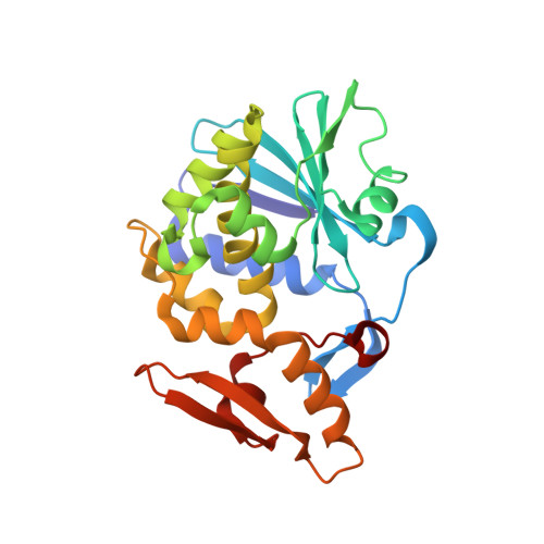 | |
UniProt | |||||
Find proteins for D9J2T9 (Momordica balsamina) Explore D9J2T9 Go to UniProtKB: D9J2T9 | |||||
Entity Groups | |||||
| Sequence Clusters | 30% Identity50% Identity70% Identity90% Identity95% Identity100% Identity | ||||
| UniProt Group | D9J2T9 | ||||
Sequence AnnotationsExpand | |||||
| |||||
Small Molecules
| Ligands 3 Unique | |||||
|---|---|---|---|---|---|
| ID | Chains | Name / Formula / InChI Key | 2D Diagram | 3D Interactions | |
| NAG Query on NAG | B [auth A] | 2-acetamido-2-deoxy-beta-D-glucopyranose C8 H15 N O6 OVRNDRQMDRJTHS-FMDGEEDCSA-N |  | ||
| ADE Query on ADE | C [auth A] | ADENINE C5 H5 N5 GFFGJBXGBJISGV-UHFFFAOYSA-N |  | ||
| GOL Query on GOL | D [auth A] | GLYCEROL C3 H8 O3 PEDCQBHIVMGVHV-UHFFFAOYSA-N |  | ||
Experimental Data & Validation
Experimental Data
- Method: X-RAY DIFFRACTION
- Resolution: 1.70 Å
- R-Value Free: 0.209
- R-Value Work: 0.175
- R-Value Observed: 0.176
- Space Group: H 3
Unit Cell:
| Length ( Å ) | Angle ( ˚ ) |
|---|---|
| a = 130.207 | α = 90 |
| b = 130.207 | β = 90 |
| c = 39.831 | γ = 120 |
| Software Name | Purpose |
|---|---|
| HKL-2000 | data collection |
| AMoRE | phasing |
| REFMAC | refinement |
| DENZO | data reduction |
| SCALEPACK | data scaling |
Entry History
Deposition Data
- Released Date: 2011-12-07 Deposition Author(s): Pandey, N., Kushwaha, G.S., Sinha, M., Bhushan, A., Kaur, P., Sharma, S., Singh, T.P.
Revision History (Full details and data files)
- Version 1.0: 2011-12-07
Type: Initial release - Version 1.1: 2012-03-28
Changes: Database references - Version 1.2: 2020-07-29
Type: Remediation
Reason: Carbohydrate remediation
Changes: Data collection, Derived calculations, Structure summary - Version 1.3: 2023-11-01
Changes: Data collection, Database references, Refinement description, Structure summary
