3U3W

Crystal Structure of Bacillus thuringiensis PlcR in complex with the peptide PapR7 and DNA


Experimental Data Snapshot

  • Method: X-RAY DIFFRACTION
  • Resolution: 2.40 Å
  • R-Value Free: 0.235 
  • R-Value Work: 0.179 
  • R-Value Observed: 0.182 

wwPDB Validation   3D Report Full Report


This is version 1.2 of the entry. See complete history


Literature

Structural basis for the activation mechanism of the PlcR virulence regulator by the quorum-sensing signal peptide PapR.

Grenha, R.Slamti, L.Nicaise, M.Refes, Y.Lereclus, D.Nessler, S.

(2013) Proc Natl Acad Sci U S A 110: 1047-1052

  • DOI: https://doi.org/10.1073/pnas.1213770110
  • Primary Citation of Related Structures:  
    3U3W, 4FSC

  • PubMed Abstract: 

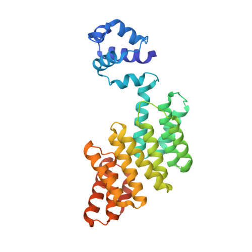
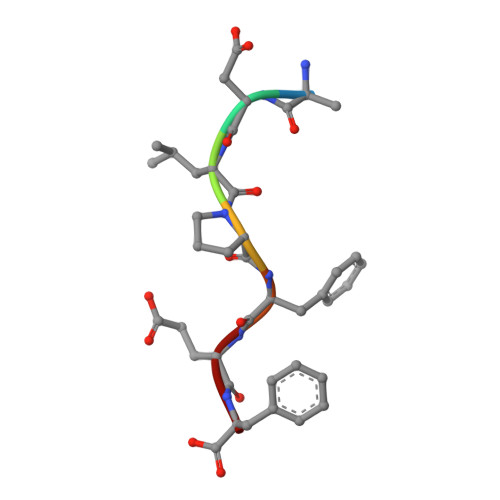
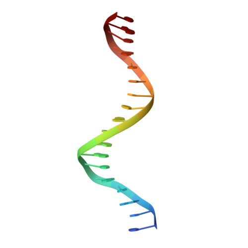
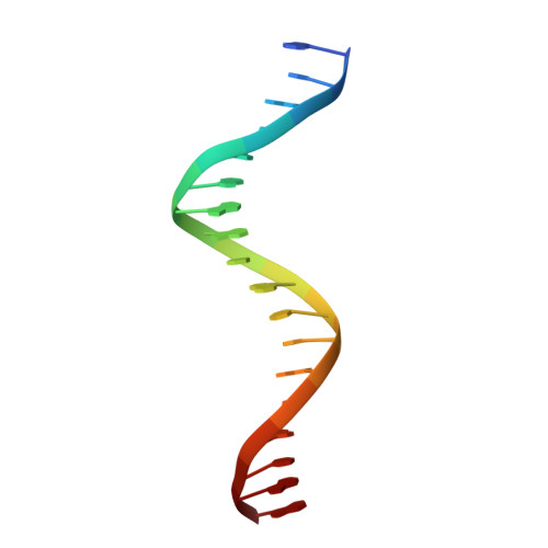
    The quorum-sensing regulator PlcR is the master regulator of most known virulence factors in Bacillus cereus. It is a helix-turn-helix (HTH)-type transcription factor activated upon binding of its cognate signaling peptide PapR on a tetratricopeptide repeat-type regulatory domain. The structural and functional properties of PlcR have defined a new family of sensor regulators, called the RNPP family (for Rap, NprR, PrgX, and PlcR), in Gram-positive bacteria. To fully understand the activation mechanism of PlcR, we took a closer look at the conformation changes induced upon binding of PapR and of its target DNA, known as PlcR-box. For that purpose we have determined the structures of the apoform of PlcR (Apo PlcR) and of the ternary complex of PlcR with PapR and the PlcR-box from the plcA promoter. Comparison of the apoform of PlcR with the previously published structure of the PlcR-PapR binary complex shows how a small conformational change induced in the C-terminal region of the tetratricopeptide repeat (TPR) domain upon peptide binding propagates via the linker helix to the N-terminal HTH DNA-binding domain. Further comparison with the PlcR-PapR-DNA ternary complex shows how the activation of the PlcR dimer allows the linker helix to undergo a drastic conformational change and subsequent proper positioning of the HTH domains in the major groove of the two half sites of the pseudopalindromic PlcR-box. Together with random mutagenesis experiments and interaction measurements using peptides from distinct pherogroups, this structural analysis allows us to propose a molecular mechanism for this functional switch.


  • Organizational Affiliation

    Laboratoire d'Enzymologie et Biochimie Structurales, Unité Propre de Recherche 3082, Centre National de la Recherche Scientifique, 91198 Gif sur Yvette, France.


Macromolecules

Find similar proteins by:  (by identity cutoff)  |  3D Structure
Entity ID: 1
MoleculeChains Sequence LengthOrganismDetailsImage
Transcriptional activator PlcR protein
A, B
293Bacillus thuringiensis Bt407Mutation(s): 0 
Gene Names: plcR
UniProt
Find proteins for Q45782 (Bacillus thuringiensis)
Explore Q45782 
Go to UniProtKB:  Q45782
Entity Groups  
Sequence Clusters30% Identity50% Identity70% Identity90% Identity95% Identity100% Identity
UniProt GroupQ45782
Sequence Annotations
Expand
  • Reference Sequence

Find similar proteins by:  Sequence   |   3D Structure  

Entity ID: 2
MoleculeChains Sequence LengthOrganismDetailsImage
C-terminus heptapeptide from PapR proteinC [auth P],
D [auth Q]
7Bacillus cereusMutation(s): 0 
UniProt
Find proteins for B7IR02 (Bacillus cereus (strain G9842))
Explore B7IR02 
Go to UniProtKB:  B7IR02
Entity Groups  
UniProt GroupB7IR02
Sequence Annotations
Expand
  • Reference Sequence

Find similar nucleic acids by:  Sequence   |   3D Structure  

Entity ID: 3
MoleculeChains LengthOrganismImage
5'-D(P*CP*TP*AP*TP*GP*CP*AP*AP*TP*AP*TP*TP*TP*CP*AP*TP*AP*T)-3'E [auth Y]18N/A
Sequence Annotations
Expand
  • Reference Sequence

Find similar nucleic acids by:  Sequence   |   3D Structure  

Entity ID: 4
MoleculeChains LengthOrganismImage
5'-D(P*AP*TP*AP*TP*GP*AP*AP*AP*TP*AP*TP*TP*GP*CP*AP*TP*AP*G)-3'F [auth Z]18N/A
Sequence Annotations
Expand
  • Reference Sequence
Experimental Data & Validation

Experimental Data

  • Method: X-RAY DIFFRACTION
  • Resolution: 2.40 Å
  • R-Value Free: 0.235 
  • R-Value Work: 0.179 
  • R-Value Observed: 0.182 
  • Space Group: P 1 21 1
Unit Cell:
Length ( Å )Angle ( ˚ )
a = 88.88α = 90
b = 71.088β = 115.53
c = 88.853γ = 90
Software Package:
Software NamePurpose
MxCuBEdata collection
PHASERphasing
PHENIXrefinement
XDSdata reduction
SCALAdata scaling

Structure Validation

View Full Validation Report


Entry History 

Deposition Data

Revision History  (Full details and data files)

  • Version 1.0: 2013-01-09
    Type: Initial release
  • Version 1.1: 2013-03-13
    Changes: Database references
  • Version 1.2: 2023-09-13
    Changes: Data collection, Database references, Derived calculations, Refinement description