3TWO

The crystal structure of CAD from Helicobacter pylori complexed with NADP(H)


Experimental Data Snapshot

  • Method: X-RAY DIFFRACTION
  • Resolution: 2.18 Å
  • R-Value Free: 0.226 
  • R-Value Work: 0.173 
  • R-Value Observed: 0.176 

wwPDB Validation   3D Report Full Report


Ligand Structure Quality Assessment 


This is version 1.2 of the entry. See complete history


Literature

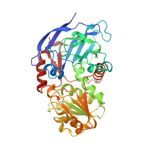
Unusual NADPH conformation in the crystal structure of a cinnamyl alcohol dehydrogenase from Helicobacter pylori in complex with NADP(H) and substrate docking analysis

Seo, K.H.Zhuang, N.N.Chen, C.Song, J.Y.Kang, H.L.Rhee, K.H.Lee, K.H.

(2012) FEBS Lett 586: 337-343

  • DOI: https://doi.org/10.1016/j.febslet.2012.01.020
  • Primary Citation of Related Structures:  
    3TWO

  • PubMed Abstract: 

    Cinnamyl alcohol dehydrogenase is a zinc- and NADPH-dependent dehydrogenase catalyzing the reversible conversion of p-hydroxycinnamaldehydes to their corresponding hydroxycinnamyl alcohols. A CAD homolog from Helicobacter pylori (HpCAD) possesses broad substrate specificities like the plant CADs and additionally a dismutation activity converting benzaldehyde to benzyl alcohol and benzoic acid. We have determined the crystal structure of HpCAD complexed with NADP(H) at 2.18Å resolution to get a better understanding of this class of CAD outside of plants. The structure of HpCAD is highly homologous to the sinapyl alcohol dehydrogenase and the plant CAD with well-conserved residues involved in catalysis and zinc binding. However, the NADP(H) binding mode of the HpCAD has been found to be significantly different from those of plant CADs.


  • Organizational Affiliation

    Division of Applied Life Science (BK21 Program), Gyeongsang National University, Jinju, Republic of Korea.


Macromolecules
Find similar proteins by:  (by identity cutoff)  |  3D Structure
Entity ID: 1
MoleculeChains Sequence LengthOrganismDetailsImage
Mannitol dehydrogenase
A, B
348Helicobacter pylori 51Mutation(s): 0 
Gene Names: cad
EC: 1.1.1.195
Entity Groups  
Sequence Clusters30% Identity50% Identity70% Identity90% Identity95% Identity100% Identity
Sequence Annotations
Expand
  • Reference Sequence
Experimental Data & Validation

Experimental Data

  • Method: X-RAY DIFFRACTION
  • Resolution: 2.18 Å
  • R-Value Free: 0.226 
  • R-Value Work: 0.173 
  • R-Value Observed: 0.176 
  • Space Group: P 21 21 21
Unit Cell:
Length ( Å )Angle ( ˚ )
a = 85.41α = 90
b = 85.68β = 90
c = 100.491γ = 90
Software Package:
Software NamePurpose
HKL-2000data collection
AMoREphasing
REFMACrefinement
HKL-2000data reduction
HKL-2000data scaling

Structure Validation

View Full Validation Report


Ligand Structure Quality Assessment 


Entry History 

Deposition Data

Revision History  (Full details and data files)

  • Version 1.0: 2011-10-12
    Type: Initial release
  • Version 1.1: 2012-05-30
    Changes: Database references
  • Version 1.2: 2023-11-01
    Changes: Data collection, Database references, Derived calculations, Refinement description