3TW4

Crystal Structure of Human Septin 7 GTPase Domain


Experimental Data Snapshot

  • Method: X-RAY DIFFRACTION
  • Resolution: 3.35 Å
  • R-Value Free: 0.365 
  • R-Value Work: 0.289 
  • R-Value Observed: 0.293 

wwPDB Validation   3D Report Full Report


Ligand Structure Quality Assessment 


This is version 1.3 of the entry. See complete history


Literature

Promiscuous interactions of human septins: The GTP binding domain of SEPT7 forms filaments within the crystal.

Serrao, V.H.Alessandro, F.Caldas, V.E.Marcal, R.L.D'Muniz Pereira, H.Thiemann, O.H.Garratt, R.C.

(2011) FEBS Lett 585: 3868-3873

  • DOI: https://doi.org/10.1016/j.febslet.2011.10.043
  • Primary Citation of Related Structures:  
    3TW4

  • PubMed Abstract: 

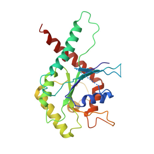
    We describe the purification, crystallization and structure for the GTP-binding domain of human septin 7 (SEPT7G). We show that it forms filaments within the crystal lattice which employ both the G and NC interfaces, similar to those seen in the hetero-filament of SEPT2/6/7. The NC interface is considered promiscuous as it is absent from the hetero-filament. Such promiscuity could provide the potential for permuting monomers along a filament in order to generate diversity in hetero-polymers. On the other hand, our results suggest that the G and NC interfaces may be necessary but insufficient for determining correct hetero-filament assembly.


  • Organizational Affiliation

    Centro de Biotecnologia Molecular Estrutural, Instituto de Física de São Carlos, Universidade de São Paulo, São Carlos, SP, Brazil.


Macromolecules
Find similar proteins by:  (by identity cutoff)  |  3D Structure
Entity ID: 1
MoleculeChains Sequence LengthOrganismDetailsImage
Septin-7
A, B
271Homo sapiensMutation(s): 0 
Gene Names: SEPT7CDC10
UniProt & NIH Common Fund Data Resources
Find proteins for Q16181 (Homo sapiens)
Explore Q16181 
Go to UniProtKB:  Q16181
PHAROS:  Q16181
GTEx:  ENSG00000122545 
Entity Groups  
Sequence Clusters30% Identity50% Identity70% Identity90% Identity95% Identity100% Identity
UniProt GroupQ16181
Sequence Annotations
Expand
  • Reference Sequence
Small Molecules
Experimental Data & Validation

Experimental Data

  • Method: X-RAY DIFFRACTION
  • Resolution: 3.35 Å
  • R-Value Free: 0.365 
  • R-Value Work: 0.289 
  • R-Value Observed: 0.293 
  • Space Group: P 61
Unit Cell:
Length ( Å )Angle ( ˚ )
a = 82.51α = 90
b = 82.51β = 90
c = 210.91γ = 120
Software Package:
Software NamePurpose
MOSFLMdata reduction
XSCALEdata scaling
PHASERphasing
DMphasing
PHENIXrefinement
PDB_EXTRACTdata extraction
DNAdata collection
XDSdata reduction

Structure Validation

View Full Validation Report


Ligand Structure Quality Assessment 


Entry History 

Deposition Data

Revision History  (Full details and data files)

  • Version 1.0: 2011-11-23
    Type: Initial release
  • Version 1.1: 2011-12-21
    Changes: Database references
  • Version 1.2: 2018-01-24
    Changes: Advisory, Structure summary
  • Version 1.3: 2023-09-13
    Changes: Advisory, Data collection, Database references, Derived calculations, Refinement description