3TV8

Pharmacological Chaperoning in Human alpha-Galactosidase


Experimental Data Snapshot

  • Method: X-RAY DIFFRACTION
  • Resolution: 2.64 Å
  • R-Value Free: 0.239 
  • R-Value Work: 0.201 
  • R-Value Observed: 0.203 

wwPDB Validation   3D Report Full Report


Ligand Structure Quality Assessment 


This is version 2.1 of the entry. See complete history


Literature

The molecular basis of pharmacological chaperoning in human alpha-galactosidase

Guce, A.I.Clark, N.E.Rogich, J.J.Garman, S.C.

(2011) Chem Biol 18: 1521-1526

  • DOI: https://doi.org/10.1016/j.chembiol.2011.10.012
  • Primary Citation of Related Structures:  
    3S5Y, 3S5Z, 3TV8

  • PubMed Abstract: 

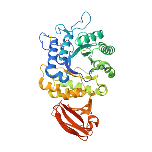
    Fabry disease patients show a deficiency in the activity of the lysosomal enzyme α-galactosidase (α-GAL or α-Gal A). One proposed treatment for Fabry disease is pharmacological chaperone therapy, where a small molecule stabilizes the α-GAL protein, leading to increased enzymatic activity. Using enzyme kinetics, tryptophan fluorescence, circular dichroism, and proteolysis assays, we show that the pharmacological chaperones 1-deoxygalactonojirimycin (DGJ) and galactose stabilize the human α-GAL glycoprotein. Crystal structures of complexes of α-GAL and chaperones explain the molecular basis for the higher potency of DGJ over galactose. Using site-directed mutagenesis, we show the higher potency of DGJ results from an ionic interaction with D170. We propose that protonation of D170 in acidic conditions leads to weaker binding of DGJ. The results establish a biochemical basis for pharmacological chaperone therapy applicable to other protein misfolding diseases.


  • Organizational Affiliation

    Department of Chemistry, University of Massachusetts Amherst, Amherst, MA 01003, USA.


Macromolecules
Find similar proteins by:  (by identity cutoff)  |  3D Structure
Entity ID: 1
MoleculeChains Sequence LengthOrganismDetailsImage
Alpha-galactosidase A
A, B
404Homo sapiensMutation(s): 1 
Gene Names: GLA
EC: 3.2.1.22
UniProt & NIH Common Fund Data Resources
Find proteins for P06280 (Homo sapiens)
Explore P06280 
Go to UniProtKB:  P06280
PHAROS:  P06280
GTEx:  ENSG00000102393 
Entity Groups  
Sequence Clusters30% Identity50% Identity70% Identity90% Identity95% Identity100% Identity
UniProt GroupP06280
Sequence Annotations
Expand
  • Reference Sequence
Oligosaccharides

Help

Entity ID: 2
MoleculeChains Length2D Diagram Glycosylation3D Interactions
beta-D-mannopyranose-(1-4)-2-acetamido-2-deoxy-beta-D-glucopyranose-(1-4)-2-acetamido-2-deoxy-beta-D-glucopyranose
C
3N-Glycosylation
Glycosylation Resources
GlyTouCan:  G15407YE
GlyCosmos:  G15407YE
GlyGen:  G15407YE
Entity ID: 3
MoleculeChains Length2D Diagram Glycosylation3D Interactions
alpha-D-mannopyranose-(1-3)-beta-D-mannopyranose-(1-4)-2-acetamido-2-deoxy-beta-D-glucopyranose-(1-4)-2-acetamido-2-deoxy-beta-D-glucopyranose
D
4N-Glycosylation
Glycosylation Resources
GlyTouCan:  G81315DD
GlyCosmos:  G81315DD
GlyGen:  G81315DD
Small Molecules
Ligands 4 Unique
IDChains Name / Formula / InChI Key2D Diagram3D Interactions
2PE
Query on 2PE

Download Ideal Coordinates CCD File 
H [auth A],
I [auth A],
O [auth B]
NONAETHYLENE GLYCOL
C18 H38 O10
YZUUTMGDONTGTN-UHFFFAOYSA-N
NAG
Query on NAG

Download Ideal Coordinates CCD File 
E [auth A],
F [auth A],
K [auth B],
L [auth B]
2-acetamido-2-deoxy-beta-D-glucopyranose
C8 H15 N O6
OVRNDRQMDRJTHS-FMDGEEDCSA-N
DGJ
Query on DGJ

Download Ideal Coordinates CCD File 
J [auth A],
P [auth B]
(2R,3S,4R,5S)-2-(hydroxymethyl)piperidine-3,4,5-triol
C6 H13 N O4
LXBIFEVIBLOUGU-DPYQTVNSSA-N
SO4
Query on SO4

Download Ideal Coordinates CCD File 
G [auth A],
M [auth B],
N [auth B]
SULFATE ION
O4 S
QAOWNCQODCNURD-UHFFFAOYSA-L
Binding Affinity Annotations 
IDSourceBinding Affinity
DGJ BindingDB:  3TV8 IC50: min: 13, max: 70 (nM) from 3 assay(s)
PDBBind:  3TV8 IC50: 70 (nM) from 1 assay(s)
Experimental Data & Validation

Experimental Data

  • Method: X-RAY DIFFRACTION
  • Resolution: 2.64 Å
  • R-Value Free: 0.239 
  • R-Value Work: 0.201 
  • R-Value Observed: 0.203 
  • Space Group: P 32 2 1
Unit Cell:
Length ( Å )Angle ( ˚ )
a = 90.465α = 90
b = 90.465β = 90
c = 216.544γ = 120
Software Package:
Software NamePurpose
SCALEPACKdata scaling
REFMACrefinement
PDB_EXTRACTdata extraction
HKL-2000data collection
DENZOdata reduction

Structure Validation

View Full Validation Report


Ligand Structure Quality Assessment 


Entry History 

Deposition Data

Revision History  (Full details and data files)

  • Version 1.0: 2012-01-04
    Type: Initial release
  • Version 2.0: 2020-07-29
    Type: Remediation
    Reason: Carbohydrate remediation
    Changes: Advisory, Atomic model, Data collection, Database references, Derived calculations, Refinement description, Structure summary
  • Version 2.1: 2023-09-13
    Changes: Data collection, Database references, Refinement description, Structure summary