3TPQ

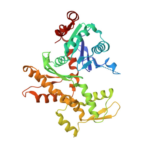
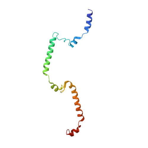
Crystal structure of wild-type MAL RPEL domain in complex with five G-actins


Experimental Data Snapshot

  • Method: X-RAY DIFFRACTION
  • Resolution: 3.45 Å
  • R-Value Free: 0.273 
  • R-Value Work: 0.224 
  • R-Value Observed: 0.227 

wwPDB Validation   3D Report Full Report


Ligand Structure Quality Assessment 


This is version 1.2 of the entry. See complete history


Literature

Sensing actin dynamics: structural basis for G-actin-sensitive nuclear import of MAL

Hirano, H.Matsuura, Y.

(2011) Biochem Biophys Res Commun 414: 373-378

  • DOI: https://doi.org/10.1016/j.bbrc.2011.09.079
  • Primary Citation of Related Structures:  
    3TPM, 3TPO, 3TPQ

  • PubMed Abstract: 

    The coordination of cytoskeletal actin dynamics with gene expression reprogramming is emerging as a crucial mechanism to control diverse cellular processes, including cell migration, differentiation and neuronal circuit assembly. The actin-binding transcriptional coactivator MAL (also known as MRTF-A/MKL1/BSAC) senses G-actin concentration and transduces Rho GTPase signals to serum response factor (SRF). MAL rapidly shuttles between the cytoplasm and the nucleus in unstimulated cells but Rho-induced depletion of G-actin leads to MAL nuclear accumulation and activation of transcription of SRF:MAL-target genes. Although the molecular and structural basis of actin-regulated nucleocytoplasmic shuttling of MAL is not understood fully, it is proposed that nuclear import of MAL is mediated by importin α/β heterodimer, and that G-actin competes with importin α/β for the binding to MAL. Here we present structural, biochemical and cell biological evidence that MAL has a classical bipartite nuclear localization signal (NLS) in the N-terminal 'RPEL' domain containing Arg-Pro-X-X-X-Glu-Leu (RPEL) motifs. The NLS residues of MAL adopt an extended conformation and bind along the surface groove of importin-α, interacting with the major- and minor-NLS binding sites. We also present a crystal structure of wild-type MAL RPEL domain in complex with five G-actins. Comparison of the importin-α- and actin-complexes revealed that the binding of G-actins to MAL is associated with folding of NLS residues into a helical conformation that is inappropriate for importin-α recognition.


  • Organizational Affiliation

    Structural Biology Research Center, Graduate School of Science, Nagoya University, Nagoya 464-8602, Japan.


Macromolecules
Find similar proteins by:  (by identity cutoff)  |  3D Structure
Entity ID: 1
MoleculeChains Sequence LengthOrganismDetailsImage
Actin, alpha skeletal muscle
A, B, C, D, E
375Oryctolagus cuniculusMutation(s): 0 
UniProt
Find proteins for P68135 (Oryctolagus cuniculus)
Explore P68135 
Go to UniProtKB:  P68135
Entity Groups  
Sequence Clusters30% Identity50% Identity70% Identity90% Identity95% Identity100% Identity
UniProt GroupP68135
Sequence Annotations
Expand
  • Reference Sequence
Find similar proteins by:  (by identity cutoff)  |  3D Structure
Entity ID: 2
MoleculeChains Sequence LengthOrganismDetailsImage
MALF [auth M]117Mus musculusMutation(s): 0 
Gene Names: Mkl1
Entity Groups  
Sequence Clusters30% Identity50% Identity70% Identity90% Identity95% Identity100% Identity
Sequence Annotations
Expand
  • Reference Sequence
Small Molecules
Ligands 2 Unique
IDChains Name / Formula / InChI Key2D Diagram3D Interactions
ATP
Query on ATP

Download Ideal Coordinates CCD File 
G [auth A],
I [auth B],
K [auth C],
M [auth D],
O [auth E]
ADENOSINE-5'-TRIPHOSPHATE
C10 H16 N5 O13 P3
ZKHQWZAMYRWXGA-KQYNXXCUSA-N
CA
Query on CA

Download Ideal Coordinates CCD File 
H [auth A],
J [auth B],
L [auth C],
N [auth D],
P [auth E]
CALCIUM ION
Ca
BHPQYMZQTOCNFJ-UHFFFAOYSA-N
Experimental Data & Validation

Experimental Data

  • Method: X-RAY DIFFRACTION
  • Resolution: 3.45 Å
  • R-Value Free: 0.273 
  • R-Value Work: 0.224 
  • R-Value Observed: 0.227 
  • Space Group: P 65 2 2
Unit Cell:
Length ( Å )Angle ( ˚ )
a = 180.737α = 90
b = 180.737β = 90
c = 382.279γ = 120
Software Package:
Software NamePurpose
REFMACrefinement
PDB_EXTRACTdata extraction
MOSFLMdata reduction
SCALAdata scaling

Structure Validation

View Full Validation Report


Ligand Structure Quality Assessment 


Entry History 

Deposition Data

Revision History  (Full details and data files)

  • Version 1.0: 2011-10-12
    Type: Initial release
  • Version 1.1: 2013-06-26
    Changes: Database references
  • Version 1.2: 2023-11-01
    Changes: Data collection, Database references, Derived calculations, Refinement description