3TPP

Crystal structure of BACE1 complexed with an inhibitor


Experimental Data Snapshot

  • Method: X-RAY DIFFRACTION
  • Resolution: 1.60 Å
  • R-Value Free: 0.180 
  • R-Value Work: 0.154 
  • R-Value Observed: 0.155 

wwPDB Validation   3D Report Full Report


Ligand Structure Quality Assessment 


This is version 1.3 of the entry. See complete history


Literature

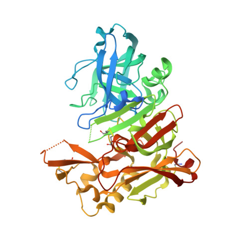
Flexibility of the flap in the active site of BACE1 as revealed by crystal structures and molecular dynamics simulations

Xu, Y.C.Li, M.J.Greenblatt, H.Chen, W.Y.Paz, A.Dym, O.Peleg, Y.Chen, T.T.Shen, X.He, J.H.Jiang, H.L.Silman, I.Sussman, J.L.

(2012) Acta Crystallogr D Biol Crystallogr 68: 13-25

  • DOI: https://doi.org/10.1107/S0907444911047251
  • Primary Citation of Related Structures:  
    3TPJ, 3TPL, 3TPP, 3TPR

  • PubMed Abstract: 

    β-Secretase (β-site amyloid precursor protein-cleaving enzyme 1; BACE1) is a transmembrane aspartic protease that cleaves the β-amyloid precursor protein en route to generation of the amyloid β-peptide (Aβ) that is believed to be responsible for the Alzheimer's disease amyloid cascade. It is thus a prime target for the development of inhibitors which may serve as drugs in the treatment and/or prevention of Alzheimer's disease. In the following determination of the crystal structures of both apo and complexed BACE1, structural analysis of all crystal structures of BACE1 deposited in the PDB and molecular dynamics (MD) simulations of monomeric and `dimeric' BACE1 were used to study conformational changes in the active-site region of the enzyme. It was observed that a flap able to cover the active site is the most flexible region, adopting multiple conformational states in the various crystal structures. Both the presence or absence of an inhibitor within the active site and the crystal packing are shown to influence the flap's conformation. An open conformation of the flap is mostly observed in the apo structures, while direct hydrogen-bonding interaction between main-chain atoms of the flap and the inhibitor is a prerequisite for the flap to adopt a closed conformation in the crystal structures of complexes. Thus, a systematic study of the conformational flexibility of the enzyme may not only contribute to structure-based drug design of BACE1 inhibitors and of other targets with flexible conformations, but may also help to better understand the mechanistic events associated with the binding of substrates and inhibitors to the enzyme.


  • Organizational Affiliation

    Drug Discovery and Design Center, Shanghai Institute of Materia Medica, Chinese Academy of Sciences, Shanghai, People's Republic of China. ycxu@mail.shcnc.ac.cn


Macromolecules
Find similar proteins by:  (by identity cutoff)  |  3D Structure
Entity ID: 1
MoleculeChains Sequence LengthOrganismDetailsImage
Beta-secretase 1433Homo sapiensMutation(s): 2 
Gene Names: BACEBACE1KIAA1149
EC: 3.4.23.46
UniProt & NIH Common Fund Data Resources
Find proteins for P56817 (Homo sapiens)
Explore P56817 
Go to UniProtKB:  P56817
PHAROS:  P56817
GTEx:  ENSG00000186318 
Entity Groups  
Sequence Clusters30% Identity50% Identity70% Identity90% Identity95% Identity100% Identity
UniProt GroupP56817
Sequence Annotations
Expand
  • Reference Sequence
Small Molecules
Ligands 4 Unique
IDChains Name / Formula / InChI Key2D Diagram3D Interactions
5HA
Query on 5HA

Download Ideal Coordinates CCD File 
B [auth A]N-[(1S,2R)-1-BENZYL-3-(CYCLOPROPYLAMINO)-2-HYDROXYPROPYL]-5-[METHYL(METHYLSULFONYL)AMINO]-N'-[(1R)-1-PHENYLETHYL]ISOPHTHALAMIDE
C31 H38 N4 O5 S
VPNIQGRFZCTBEZ-SPTGULJVSA-N
SO4
Query on SO4

Download Ideal Coordinates CCD File 
V [auth A],
W [auth A]
SULFATE ION
O4 S
QAOWNCQODCNURD-UHFFFAOYSA-L
URE
Query on URE

Download Ideal Coordinates CCD File 
K [auth A]
L [auth A]
M [auth A]
N [auth A]
O [auth A]
K [auth A],
L [auth A],
M [auth A],
N [auth A],
O [auth A],
P [auth A],
Q [auth A],
R [auth A],
S [auth A],
T [auth A],
U [auth A]
UREA
C H4 N2 O
XSQUKJJJFZCRTK-UHFFFAOYSA-N
CL
Query on CL

Download Ideal Coordinates CCD File 
C [auth A]
D [auth A]
E [auth A]
F [auth A]
G [auth A]
C [auth A],
D [auth A],
E [auth A],
F [auth A],
G [auth A],
H [auth A],
I [auth A],
J [auth A]
CHLORIDE ION
Cl
VEXZGXHMUGYJMC-UHFFFAOYSA-M
Binding Affinity Annotations 
IDSourceBinding Affinity
5HA BindingDB:  3TPP Ki: 233 (nM) from 1 assay(s)
IC50: min: 2, max: 1.50e+4 (nM) from 13 assay(s)
EC50: 43 (nM) from 1 assay(s)
Binding MOAD:  3TPP IC50: 15 (nM) from 1 assay(s)
PDBBind:  3TPP IC50: 15 (nM) from 1 assay(s)
Experimental Data & Validation

Experimental Data

  • Method: X-RAY DIFFRACTION
  • Resolution: 1.60 Å
  • R-Value Free: 0.180 
  • R-Value Work: 0.154 
  • R-Value Observed: 0.155 
  • Space Group: C 2 2 21
Unit Cell:
Length ( Å )Angle ( ˚ )
a = 104.548α = 90
b = 128.202β = 90
c = 76.535γ = 90
Software Package:
Software NamePurpose
DENZOdata reduction
SCALEPACKdata scaling
PHENIXrefinement
PDB_EXTRACTdata extraction
HKL-2000data collection
HKL-2000data reduction
HKL-2000data scaling
MOLREPphasing

Structure Validation

View Full Validation Report


Ligand Structure Quality Assessment 


Entry History 

Deposition Data

Revision History  (Full details and data files)

  • Version 1.0: 2011-11-23
    Type: Initial release
  • Version 1.1: 2012-06-13
    Changes: Database references
  • Version 1.2: 2017-11-08
    Changes: Refinement description
  • Version 1.3: 2023-11-01
    Changes: Data collection, Database references, Derived calculations, Refinement description