3TKM

Crystal structure PPAR delta binding GW0742


Experimental Data Snapshot

  • Method: X-RAY DIFFRACTION
  • Resolution: 1.95 Å
  • R-Value Free: 0.245 
  • R-Value Work: 0.197 
  • R-Value Observed: 0.199 

wwPDB Validation   3D Report Full Report


Ligand Structure Quality Assessment 


This is version 1.2 of the entry. See complete history


Literature

Structural Insights into Human Peroxisome Proliferator Activated Receptor Delta (PPAR-Delta) Selective Ligand Binding.

Batista, F.A.Trivella, D.B.Bernardes, A.Gratieri, J.Oliveira, P.S.Figueira, A.C.Webb, P.Polikarpov, I.

(2012) PLoS One 7: e33643-e33643

  • DOI: https://doi.org/10.1371/journal.pone.0033643
  • Primary Citation of Related Structures:  
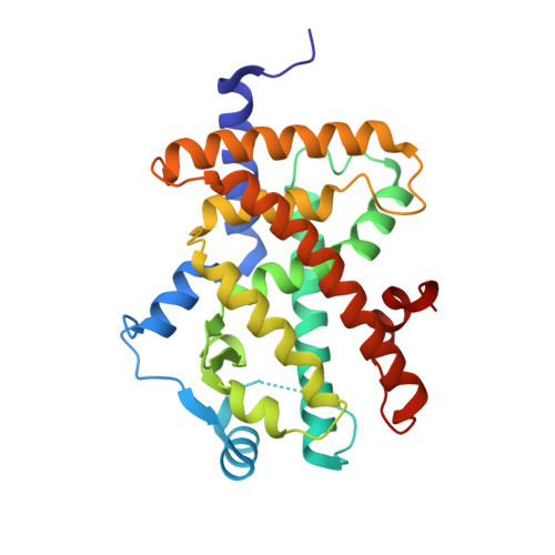
    3TKM

  • PubMed Abstract: 

    Peroxisome proliferator activated receptors (PPARs δ, α and γ) are closely related transcription factors that exert distinct effects on fatty acid and glucose metabolism, cardiac disease, inflammatory response and other processes. Several groups developed PPAR subtype specific modulators to trigger desirable effects of particular PPARs without harmful side effects associated with activation of other subtypes. Presently, however, many compounds that bind to one of the PPARs cross-react with others and rational strategies to obtain highly selective PPAR modulators are far from clear. GW0742 is a synthetic ligand that binds PPARδ more than 300-fold more tightly than PPARα or PPARγ but the structural basis of PPARδ:GW0742 interactions and reasons for strong selectivity are not clear. Here we report the crystal structure of the PPARδ:GW0742 complex. Comparisons of the PPARδ:GW0742 complex with published structures of PPARs in complex with α and γ selective agonists and pan agonists suggests that two residues (Val312 and Ile328) in the buried hormone binding pocket play special roles in PPARδ selective binding and experimental and computational analysis of effects of mutations in these residues confirms this and suggests that bulky substituents that line the PPARα and γ ligand binding pockets as structural barriers for GW0742 binding. This analysis suggests general strategies for selective PPARδ ligand design.


  • Organizational Affiliation

    Instituto de Física de São Carlos, Universidade de São Paulo, São Carlos, Sao Paulo, Brazil.


Macromolecules
Find similar proteins by:  (by identity cutoff)  |  3D Structure
Entity ID: 1
MoleculeChains Sequence LengthOrganismDetailsImage
Peroxisome proliferator-activated receptor delta275Homo sapiensMutation(s): 0 
Gene Names: PPARDNR1C2PPARB
UniProt & NIH Common Fund Data Resources
Find proteins for Q03181 (Homo sapiens)
Explore Q03181 
Go to UniProtKB:  Q03181
PHAROS:  Q03181
GTEx:  ENSG00000112033 
Entity Groups  
Sequence Clusters30% Identity50% Identity70% Identity90% Identity95% Identity100% Identity
UniProt GroupQ03181
Sequence Annotations
Expand
  • Reference Sequence
Small Molecules
Ligands 2 Unique
IDChains Name / Formula / InChI Key2D Diagram3D Interactions
GW0
Query on GW0

Download Ideal Coordinates CCD File 
B [auth A]{4-[({2-[3-fluoro-4-(trifluoromethyl)phenyl]-4-methyl-1,3-thiazol-5-yl}methyl)sulfanyl]-2-methylphenoxy}acetic acid
C21 H17 F4 N O3 S2
HWVNEWGKWRGSRK-UHFFFAOYSA-N
GOL
Query on GOL

Download Ideal Coordinates CCD File 
C [auth A]GLYCEROL
C3 H8 O3
PEDCQBHIVMGVHV-UHFFFAOYSA-N
Binding Affinity Annotations 
IDSourceBinding Affinity
GW0 Binding MOAD:  3TKM Kd: 22 (nM) from 1 assay(s)
BindingDB:  3TKM Kd: 0.4 (nM) from 1 assay(s)
IC50: 1 (nM) from 1 assay(s)
EC50: min: 1, max: 300 (nM) from 9 assay(s)
PDBBind:  3TKM IC50: 1 (nM) from 1 assay(s)
Experimental Data & Validation

Experimental Data

  • Method: X-RAY DIFFRACTION
  • Resolution: 1.95 Å
  • R-Value Free: 0.245 
  • R-Value Work: 0.197 
  • R-Value Observed: 0.199 
  • Space Group: P 21 21 21
Unit Cell:
Length ( Å )Angle ( ˚ )
a = 35.466α = 90
b = 74.766β = 90
c = 96.287γ = 90
Software Package:
Software NamePurpose
MAR345dtbdata collection
PHASERphasing
PHENIXrefinement
DENZOdata reduction
SCALEPACKdata scaling

Structure Validation

View Full Validation Report


Ligand Structure Quality Assessment 


Entry History 

Deposition Data

Revision History  (Full details and data files)

  • Version 1.0: 2012-07-04
    Type: Initial release
  • Version 1.1: 2020-10-21
    Changes: Data collection, Database references, Derived calculations
  • Version 1.2: 2023-09-13
    Changes: Data collection, Database references, Refinement description