3TJ6

human vinculin head domain (Vh1, residues 1-258) in complex with the vinculin binding site of the surface cell antigen 4 (sca4-VBS-C; residues 812-835) from Rickettsia rickettsii


Experimental Data Snapshot

  • Method: X-RAY DIFFRACTION
  • Resolution: 2.76 Å
  • R-Value Free: 0.235 
  • R-Value Work: 0.193 
  • R-Value Observed: 0.196 

wwPDB Validation   3D Report Full Report


This is version 1.4 of the entry. See complete history


Literature

The rickettsia surface cell antigen 4 applies mimicry to bind to and activate vinculin.

Park, H.Lee, J.H.Gouin, E.Cossart, P.Izard, T.

(2011) J Biol Chem 286: 35096-35103

  • DOI: https://doi.org/10.1074/jbc.M111.263855
  • Primary Citation of Related Structures:  
    3TJ5, 3TJ6

  • PubMed Abstract: 

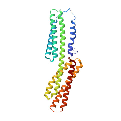
    Pathogenic Rickettsia species cause high morbidity and mortality, especially R. prowazekii, the causative agent of typhus. Like many intracellular pathogens, Rickettsia exploit the cytoskeleton to enter and spread within the host cell. Here we report that the cell surface antigen sca4 of Rickettsia co-localizes with vinculin in cells at sites of focal adhesions in sca4-transfected cells and that sca4 binds to and activates vinculin through two vinculin binding sites (VBSs) that are conserved across all Rickettsia. Remarkably, this occurs through molecular mimicry of the vinculin-talin interaction that is also seen with the IpaA invasin of the intracellular pathogen Shigella, where binding of these VBSs to the vinculin seven-helix bundle head domain (Vh1) displaces intramolecular interactions with the vinculin tail domain that normally clamp vinculin in an inactive state. Finally, the vinculin·sca4-VBS crystal structures reveal that vinculin adopts a new conformation when bound to the C-terminal VBS of sca4. Collectively, our data define the mechanism by which sca4 activates vinculin and interacts with the actin cytoskeleton, and they suggest important roles for vinculin in Rickettsia pathogenesis.


  • Organizational Affiliation

    Cell Adhesion Laboratory, Department of Cancer Biology, The Scripps Research Institute, Jupiter, Florida 33458, USA.


Macromolecules
Find similar proteins by:  (by identity cutoff)  |  3D Structure
Entity ID: 1
MoleculeChains Sequence LengthOrganismDetailsImage
Vinculin257Homo sapiensMutation(s): 0 
Gene Names: VCL
UniProt & NIH Common Fund Data Resources
Find proteins for P18206 (Homo sapiens)
Explore P18206 
Go to UniProtKB:  P18206
PHAROS:  P18206
GTEx:  ENSG00000035403 
Entity Groups  
Sequence Clusters30% Identity50% Identity70% Identity90% Identity95% Identity100% Identity
UniProt GroupP18206
Sequence Annotations
Expand
  • Reference Sequence

Find similar proteins by:  Sequence   |   3D Structure  

Entity ID: 2
MoleculeChains Sequence LengthOrganismDetailsImage
Antigenic heat-stable 120 kDa protein23Rickettsia rickettsii str. IowaMutation(s): 0 
Gene Names: RrIowa_0797
UniProt
Find proteins for B0BXR4 (Rickettsia rickettsii (strain Iowa))
Explore B0BXR4 
Go to UniProtKB:  B0BXR4
Entity Groups  
Sequence Clusters30% Identity50% Identity70% Identity90% Identity95% Identity100% Identity
UniProt GroupB0BXR4
Sequence Annotations
Expand
  • Reference Sequence
Experimental Data & Validation

Experimental Data

  • Method: X-RAY DIFFRACTION
  • Resolution: 2.76 Å
  • R-Value Free: 0.235 
  • R-Value Work: 0.193 
  • R-Value Observed: 0.196 
  • Space Group: I 21 3
Unit Cell:
Length ( Å )Angle ( ˚ )
a = 141.373α = 90
b = 141.373β = 90
c = 141.373γ = 90
Software Package:
Software NamePurpose
Blu-Icedata collection
MOLREPphasing
BUSTERrefinement
autoPROCdata scaling

Structure Validation

View Full Validation Report


Entry History 

Deposition Data

Revision History  (Full details and data files)

  • Version 1.0: 2011-09-07
    Type: Initial release
  • Version 1.1: 2011-11-02
    Changes: Database references
  • Version 1.2: 2013-05-01
    Changes: Other
  • Version 1.3: 2017-11-08
    Changes: Refinement description
  • Version 1.4: 2023-09-13
    Changes: Data collection, Database references, Refinement description