3TIW

Crystal structure of p97N in complex with the C-terminus of gp78


Experimental Data Snapshot

  • Method: X-RAY DIFFRACTION
  • Resolution: 1.80 Å
  • R-Value Free: 0.171 
  • R-Value Work: 0.147 
  • R-Value Observed: 0.148 

wwPDB Validation   3D Report Full Report


This is version 1.3 of the entry. See complete history


Literature

The Structural and Functional Basis of the p97/Valosin-containing Protein (VCP)-interacting Motif (VIM): MUTUALLY EXCLUSIVE BINDING OF COFACTORS TO THE N-TERMINAL DOMAIN OF p97.

Hanzelmann, P.Schindelin, H.

(2011) J Biol Chem 286: 38679-38690

  • DOI: https://doi.org/10.1074/jbc.M111.274506
  • Primary Citation of Related Structures:  
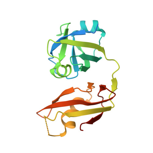
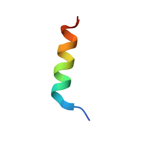
    3TIW

  • PubMed Abstract: 

    The AAA (ATPase associated with various cellular activities) ATPase p97, also referred to as valosin-containing protein (VCP), mediates essential cellular processes, including ubiquitin-dependent protein degradation, and has been linked to several human proteinopathies. p97 interacts with multiple cofactors via its N-terminal (p97N) domain, a subset of which contain the VCP-interacting motif (VIM). We have determined the crystal structure of the p97N domain in complex with the VIM of the ubiquitin E3 ligase gp78 at 1.8 Å resolution. The α-helical VIM peptide binds into a groove located in between the two subdomains of the p97N domain. Interaction studies of several VIM proteins reveal that these cofactors display dramatically different affinities, ranging from high affinity interactions characterized by dissociation constants of ∼20 nm for gp78 and ANKZF1 to only weak binding in our assays. The contribution of individual p97 residues to VIM binding was analyzed, revealing that identical substitutions do not affect all cofactors in the same way. Taken together, the biochemical and structural studies define the framework for recognition of VIM-containing cofactors by p97. Of particular interest to the regulation of p97 by its cofactors, our structure reveals that the bound α-helical peptides of VIM-containing cofactors overlap with the binding site for cofactors containing the ubiquitin regulatory X (UBX) domain present in the UBX protein family or the ubiquitin-like domain of NPL4 as further corroborated by biochemical data. These results extend the concept that competitive binding is a crucial determinant in p97-cofactor interactions.


  • Organizational Affiliation

    Rudolf Virchow Center for Experimental Biomedicine, University of Würzburg, Josef-Schneider-Strasse 2, 97080 Würzburg, Germany. Electronic address: petra.haenzelmann@virchow.uni-wuerzburg.de.


Macromolecules
Find similar proteins by:  (by identity cutoff)  |  3D Structure
Entity ID: 1
MoleculeChains Sequence LengthOrganismDetailsImage
Transitional endoplasmic reticulum ATPaseA,
C [auth B]
187Homo sapiensMutation(s): 0 
Gene Names: VCP
UniProt & NIH Common Fund Data Resources
Find proteins for P55072 (Homo sapiens)
Explore P55072 
Go to UniProtKB:  P55072
PHAROS:  P55072
GTEx:  ENSG00000165280 
Entity Groups  
Sequence Clusters30% Identity50% Identity70% Identity90% Identity95% Identity100% Identity
UniProt GroupP55072
Sequence Annotations
Expand
  • Reference Sequence

Find similar proteins by:  Sequence   |   3D Structure  

Entity ID: 2
MoleculeChains Sequence LengthOrganismDetailsImage
E3 ubiquitin-protein ligase AMFRB [auth C],
D
19Homo sapiensMutation(s): 0 
EC: 6.3.2
UniProt & NIH Common Fund Data Resources
Find proteins for Q9UKV5 (Homo sapiens)
Explore Q9UKV5 
Go to UniProtKB:  Q9UKV5
PHAROS:  Q9UKV5
GTEx:  ENSG00000159461 
Entity Groups  
Sequence Clusters30% Identity50% Identity70% Identity90% Identity95% Identity100% Identity
UniProt GroupQ9UKV5
Sequence Annotations
Expand
  • Reference Sequence
Experimental Data & Validation

Experimental Data

  • Method: X-RAY DIFFRACTION
  • Resolution: 1.80 Å
  • R-Value Free: 0.171 
  • R-Value Work: 0.147 
  • R-Value Observed: 0.148 
  • Space Group: P 32
Unit Cell:
Length ( Å )Angle ( ˚ )
a = 70.24α = 90
b = 70.24β = 90
c = 74.82γ = 120
Software Package:
Software NamePurpose
MOSFLMdata reduction
SCALAdata scaling
PHASERphasing
PHENIXrefinement
PDB_EXTRACTdata extraction
MxCuBEdata collection

Structure Validation

View Full Validation Report


Entry History 

Deposition Data

Revision History  (Full details and data files)

  • Version 1.0: 2011-09-14
    Type: Initial release
  • Version 1.1: 2011-11-02
    Changes: Database references
  • Version 1.2: 2011-11-16
    Changes: Database references
  • Version 1.3: 2023-09-13
    Changes: Data collection, Database references, Derived calculations, Refinement description