3TG5

Structure of SMYD2 in complex with p53 and SAH


Experimental Data Snapshot

  • Method: X-RAY DIFFRACTION
  • Resolution: 2.30 Å
  • R-Value Free: 0.263 
  • R-Value Work: 0.241 
  • R-Value Observed: 0.242 

wwPDB Validation   3D Report Full Report


Ligand Structure Quality Assessment 


This is version 1.2 of the entry. See complete history


Literature

Structure of human SMYD2 reveals the basis of p53 tumor suppressor methylation

Wang, L.Li, L.Zhang, H.Luo, X.Dai, J.Zhou, S.Gu, J.Zhu, J.Atadja, P.Lu, C.Li, E.Zhao, K.

(2011) J Biol Chem 

  • DOI: https://doi.org/10.1074/jbc.M111.262410
  • Primary Citation of Related Structures:  
    3TG4, 3TG5

  • PubMed Abstract: 

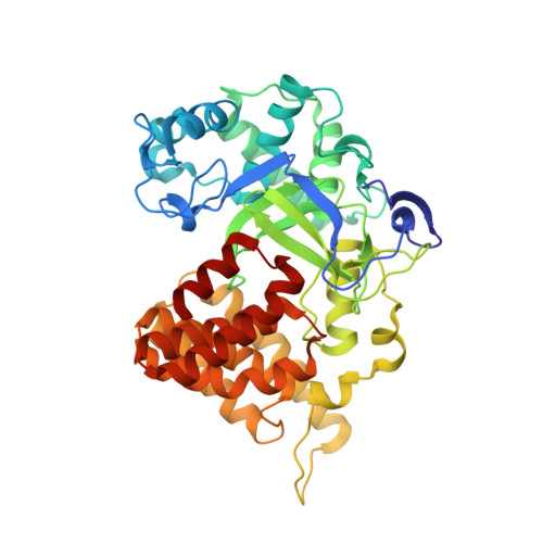
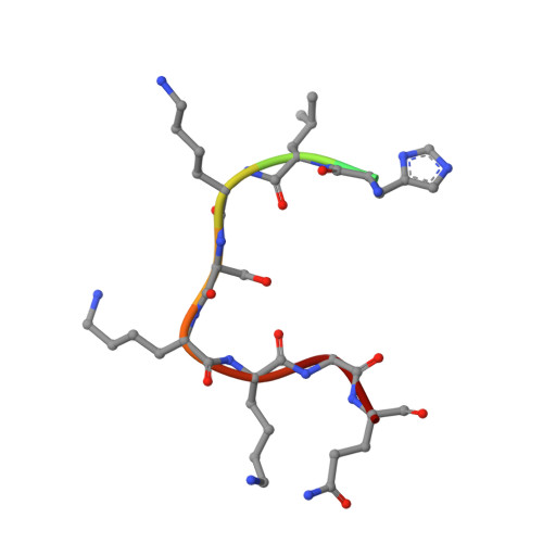
    SMYD2 belongs to a subfamily of histone lysine methyltransferase and was recently identified to methylate tumor suppressor p53 and Rb. Here we report that SMYD2 prefers to methylate p53 Lys-370 over histone substrates in vitro. Consistently, the level of endogenous p53 Lys-370 monomethylation is significantly elevated when SMYD2 is overexpressed in vivo. We have solved the high resolution crystal structures of the full-length SMYD2 protein in binary complex with its cofactor S-adenosylmethionine and in ternary complex with cofactor product S-adenosylhomocysteine and p53 substrate peptide (residues 368-375), respectively. p53 peptide binds to a deep pocket of the interface between catalytic SET(1-282) and C-terminal domain (CTD) with an unprecedented U-shaped conformation. Subtle conformational change exists around the p53 binding site between the binary and ternary structures, in particular the tetratricopeptide repeat motif of the CTD. In addition, a unique EDEE motif between the loop of anti-parallel β7 and β8 sheets of the SET core not only interacts with p53 substrate but also forms a hydrogen bond network with residues from CTD. These observations suggest that the tetratricopeptide repeat and EDEE motif may play an important role in determining p53 substrate binding specificity. This is further verified by the findings that deletion of the CTD domain drastically reduces the methylation activity of SMYD2 to p53 protein. Meanwhile, mutation of EDEE residues impairs both the binding and the enzymatic activity of SMYD2 to p53 Lys-370. These data together reveal the molecular basis of SMYD2 in specifically recognizing and regulating functions of p53 tumor suppressor through Lys-370 monomethylation.


  • Organizational Affiliation

    Novartis Institutes for BioMedical Research, Building 8, Lane 898, Halei Road, Pudong, Shanghai 201203, China.


Macromolecules
Find similar proteins by:  (by identity cutoff)  |  3D Structure
Entity ID: 1
MoleculeChains Sequence LengthOrganismDetailsImage
N-lysine methyltransferase SMYD2433Homo sapiensMutation(s): 0 
Gene Names: SMYD2KMT3C
EC: 2.1.1 (PDB Primary Data), 2.1.1.43 (PDB Primary Data)
UniProt & NIH Common Fund Data Resources
Find proteins for Q9NRG4 (Homo sapiens)
Explore Q9NRG4 
Go to UniProtKB:  Q9NRG4
PHAROS:  Q9NRG4
GTEx:  ENSG00000143499 
Entity Groups  
Sequence Clusters30% Identity50% Identity70% Identity90% Identity95% Identity100% Identity
UniProt GroupQ9NRG4
Sequence Annotations
Expand
  • Reference Sequence

Find similar proteins by:  Sequence   |   3D Structure  

Entity ID: 2
MoleculeChains Sequence LengthOrganismDetailsImage
Cellular tumor antigen p5311Homo sapiensMutation(s): 0 
UniProt & NIH Common Fund Data Resources
Find proteins for P04637 (Homo sapiens)
Explore P04637 
Go to UniProtKB:  P04637
PHAROS:  P04637
GTEx:  ENSG00000141510 
Entity Groups  
Sequence Clusters30% Identity50% Identity70% Identity90% Identity95% Identity100% Identity
UniProt GroupP04637
Sequence Annotations
Expand
  • Reference Sequence
Experimental Data & Validation

Experimental Data

  • Method: X-RAY DIFFRACTION
  • Resolution: 2.30 Å
  • R-Value Free: 0.263 
  • R-Value Work: 0.241 
  • R-Value Observed: 0.242 
  • Space Group: P 21 21 21
Unit Cell:
Length ( Å )Angle ( ˚ )
a = 53.36α = 90
b = 71.67β = 90
c = 121.25γ = 90
Software Package:
Software NamePurpose
HKL-2000data collection
MOLREPphasing
REFMACrefinement
HKL-2000data reduction
SCALAdata scaling

Structure Validation

View Full Validation Report


Ligand Structure Quality Assessment 


Entry History 

Deposition Data

Revision History  (Full details and data files)

  • Version 1.0: 2011-08-31
    Type: Initial release
  • Version 1.1: 2011-09-14
    Changes: Database references
  • Version 1.2: 2023-11-01
    Changes: Data collection, Database references, Derived calculations, Refinement description