3TDJ

Crystal structure of the GluA2 ligand-binding domain (S1S2J-L483Y-N754S) in complex with glutamate and BPAM-97 at 1.95 A resolution


Experimental Data Snapshot

  • Method: X-RAY DIFFRACTION
  • Resolution: 1.95 Å
  • R-Value Free: 0.215 
  • R-Value Work: 0.170 
  • R-Value Observed: 0.172 

wwPDB Validation   3D Report Full Report


Ligand Structure Quality Assessment 


This is version 1.3 of the entry. See complete history


Literature

Thermodynamics and structural analysis of positive allosteric modulation of the ionotropic glutamate receptor GluA2.

Krintel, C.Frydenvang, K.Olsen, L.Kristensen, M.T.de Barrios, O.Naur, P.Francotte, P.Pirotte, B.Gajhede, M.Kastrup, J.S.

(2012) Biochem J 441: 173-178

  • DOI: https://doi.org/10.1042/BJ20111221
  • Primary Citation of Related Structures:  
    3TDJ, 3TKD

  • PubMed Abstract: 

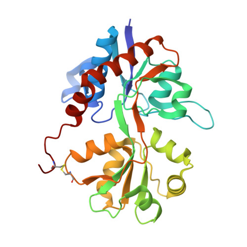
    Positive allosteric modulators of the ionotropic glutamate receptor-2 (GluA2) are promising compounds for the treatment of cognitive disorders, e.g. Alzheimer's disease. These modulators bind within the dimer interface of the LBD (ligand-binding domain) and stabilize the agonist-bound conformation slowing receptor desensitization and/or deactivation. In the present study, we employ isothermal titration calorimetry to determine binding affinities and thermodynamic details of binding of modulators of GluA2. A mutant of the LBD of GluA2 (LBD-L483Y-N754S) that forms a stable dimer in solution was used. The potent GluA2 modulator BPAM-97 was used as a reference compound. Evidence that BPAM-97 binds in the same pocket as the well-known GluA2 modulator cyclothiazide was obtained from X-ray structures. The LBD-L483Y-N754S:BPAM-97 complex has a Kd of 5.6 μM (ΔH=-4.9 kcal/mol, -TΔS=-2.3 kcal/mol; where 1 kcal≈4.187 kJ). BPAM-97 was used in a displacement assay to determine a Kd of 0.46 mM (ΔH=-1.2 kcal/mol, -TΔS=-3.3 kcal/mol) for the LBD-L483Y-N754S:IDRA-21 complex. The major structural factors increasing the potency of BPAM-97 over IDRA-21 are the increased van der Waals contacts to, primarily, Met496 in GluA2 imposed by the ethyl substituent of BPAM-97. These results add important information on binding affinities and thermodynamic details, and provide a new tool in the development of drugs against cognitive disorders.


  • Organizational Affiliation

    Department of Medicinal Chemistry, Faculty of Pharmaceutical Sciences, University of Copenhagen, Universitetsparken 2, DK-2100 Copenhagen, Denmark.


Macromolecules
Find similar proteins by:  (by identity cutoff)  |  3D Structure
Entity ID: 1
MoleculeChains Sequence LengthOrganismDetailsImage
Glutamate receptor 2
A, B
263Rattus norvegicusMutation(s): 2 
Gene Names: Glur2Gria2
UniProt
Find proteins for P19491 (Rattus norvegicus)
Explore P19491 
Go to UniProtKB:  P19491
Entity Groups  
Sequence Clusters30% Identity50% Identity70% Identity90% Identity95% Identity100% Identity
UniProt GroupP19491
Sequence Annotations
Expand
  • Reference Sequence
Small Molecules
Ligands 5 Unique
IDChains Name / Formula / InChI Key2D Diagram3D Interactions
3TJ
Query on 3TJ

Download Ideal Coordinates CCD File 
N [auth B],
O [auth B]
4-ethyl-7-fluoro-3,4-dihydro-2H-1,2,4-benzothiadiazine 1,1-dioxide
C9 H11 F N2 O2 S
MALNTEMXARBOND-UHFFFAOYSA-N
GLU
Query on GLU

Download Ideal Coordinates CCD File 
C [auth A],
P [auth B]
GLUTAMIC ACID
C5 H9 N O4
WHUUTDBJXJRKMK-VKHMYHEASA-N
SO4
Query on SO4

Download Ideal Coordinates CCD File 
D [auth A],
Q [auth B],
X [auth B]
SULFATE ION
O4 S
QAOWNCQODCNURD-UHFFFAOYSA-L
GOL
Query on GOL

Download Ideal Coordinates CCD File 
G [auth A]
H [auth A]
I [auth A]
J [auth A]
K [auth A]
G [auth A],
H [auth A],
I [auth A],
J [auth A],
K [auth A],
R [auth B],
S [auth B],
T [auth B],
U [auth B],
V [auth B],
W [auth B]
GLYCEROL
C3 H8 O3
PEDCQBHIVMGVHV-UHFFFAOYSA-N
CL
Query on CL

Download Ideal Coordinates CCD File 
E [auth A],
F [auth A],
L [auth A],
M [auth A],
Y [auth B]
CHLORIDE ION
Cl
VEXZGXHMUGYJMC-UHFFFAOYSA-M
Binding Affinity Annotations 
IDSourceBinding Affinity
3TJ PDBBind:  3TDJ Kd: 5600 (nM) from 1 assay(s)
BindingDB:  3TDJ Kd: 5600 (nM) from 1 assay(s)
Experimental Data & Validation

Experimental Data

  • Method: X-RAY DIFFRACTION
  • Resolution: 1.95 Å
  • R-Value Free: 0.215 
  • R-Value Work: 0.170 
  • R-Value Observed: 0.172 
  • Space Group: P 21 21 2
Unit Cell:
Length ( Å )Angle ( ˚ )
a = 98.86α = 90
b = 121.3β = 90
c = 47.31γ = 90
Software Package:
Software NamePurpose
PHASERphasing
PHENIXrefinement
MOSFLMdata reduction
SCALAdata scaling

Structure Validation

View Full Validation Report


Ligand Structure Quality Assessment 


Entry History 

Deposition Data

Revision History  (Full details and data files)

  • Version 1.0: 2011-09-21
    Type: Initial release
  • Version 1.1: 2011-12-28
    Changes: Database references
  • Version 1.2: 2017-08-09
    Changes: Data collection, Refinement description, Source and taxonomy
  • Version 1.3: 2023-09-13
    Changes: Data collection, Database references, Derived calculations, Refinement description