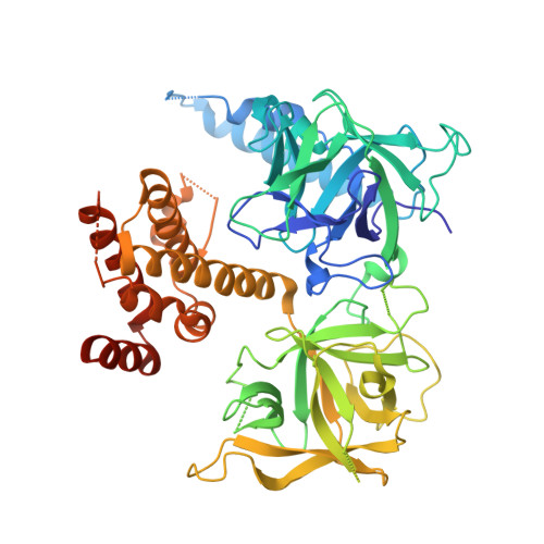
3T8S

Apo and InsP3-bound Crystal Structures of the Ligand-Binding Domain of an InsP3 Receptor


Experimental Data Snapshot

  • Method: X-RAY DIFFRACTION
  • Resolution: 3.77 Å
  • R-Value Free: 0.292 
  • R-Value Work: 0.277 
  • R-Value Observed: 0.278 

wwPDB Validation   3D Report Full Report


Ligand Structure Quality Assessment 


This is version 1.3 of the entry. See complete history


Literature

Apo and InsP(3)-bound crystal structures of the ligand-binding domain of an InsP(3) receptor.

Lin, C.C.Baek, K.Lu, Z.

(2011) Nat Struct Mol Biol 18: 1172-1174

  • DOI: https://doi.org/10.1038/nsmb.2112
  • Primary Citation of Related Structures:  
    3T8S

  • PubMed Abstract: 

    We report the crystal structures of the ligand-binding domain (LBD) of a rat inositol 1,4,5-trisphosphate receptor (InsP(3)R) in its apo and InsP(3)-bound conformations. Comparison of these two conformations reveals that LBD's first β-trefoil fold (β-TF1) and armadillo repeat fold (ARF) move together as a unit relative to its second β-trefoil fold (β-TF2). Whereas apo LBD may spontaneously transition between gating conformations, InsP(3) binding shifts this equilibrium toward the active state.


  • Organizational Affiliation

    Department of Physiology, Howard Hughes Medical Institute, Perelman School of Medicine, University of Pennsylvania, Philadelphia, Pennsylvania, USA.


Macromolecules
Find similar proteins by:  (by identity cutoff)  |  3D Structure
Entity ID: 1
MoleculeChains Sequence LengthOrganismDetailsImage
Inositol 1,4,5-trisphosphate receptor type 1
A, B
585Rattus norvegicusMutation(s): 0 
Gene Names: Insp3rItpr1
UniProt
Find proteins for P29994 (Rattus norvegicus)
Explore P29994 
Go to UniProtKB:  P29994
Entity Groups  
Sequence Clusters30% Identity50% Identity70% Identity90% Identity95% Identity100% Identity
UniProt GroupP29994
Sequence Annotations
Expand
  • Reference Sequence
Small Molecules
Ligands 1 Unique
IDChains Name / Formula / InChI Key2D Diagram3D Interactions
I3P
Query on I3P

Download Ideal Coordinates CCD File 
C [auth B]D-MYO-INOSITOL-1,4,5-TRIPHOSPHATE
C6 H15 O15 P3
MMWCIQZXVOZEGG-XJTPDSDZSA-N
Binding Affinity Annotations 
IDSourceBinding Affinity
I3P PDBBind:  3T8S Kd: 2.82 (nM) from 1 assay(s)
BindingDB:  3T8S Kd: min: 0.21, max: 260 (nM) from 5 assay(s)
IC50: 300 (nM) from 1 assay(s)
EC50: min: 20, max: 209 (nM) from 7 assay(s)
Experimental Data & Validation

Experimental Data

  • Method: X-RAY DIFFRACTION
  • Resolution: 3.77 Å
  • R-Value Free: 0.292 
  • R-Value Work: 0.277 
  • R-Value Observed: 0.278 
  • Space Group: C 1 2 1
Unit Cell:
Length ( Å )Angle ( ˚ )
a = 159.829α = 90
b = 84.877β = 116.92
c = 95.09γ = 90
Software Package:
Software NamePurpose
DENZOdata reduction
SCALEPACKdata scaling
PHASERphasing
REFMACrefinement
PDB_EXTRACTdata extraction
HKL-2000data collection

Structure Validation

View Full Validation Report


Ligand Structure Quality Assessment 


Entry History 

Deposition Data

Revision History  (Full details and data files)

  • Version 1.0: 2011-09-07
    Type: Initial release
  • Version 1.1: 2011-09-21
    Changes: Database references
  • Version 1.2: 2011-10-26
    Changes: Database references
  • Version 1.3: 2023-09-13
    Changes: Data collection, Database references, Derived calculations, Refinement description