3T6B

Structure of human DPPIII in complex with the opioid peptide Tynorphin, at 2.4 Angstroms


Experimental Data Snapshot

  • Method: X-RAY DIFFRACTION
  • Resolution: 2.40 Å
  • R-Value Free: 0.258 
  • R-Value Work: 0.197 
  • R-Value Observed: 0.200 

wwPDB Validation   3D Report Full Report


This is version 1.5 of the entry. See complete history


Literature

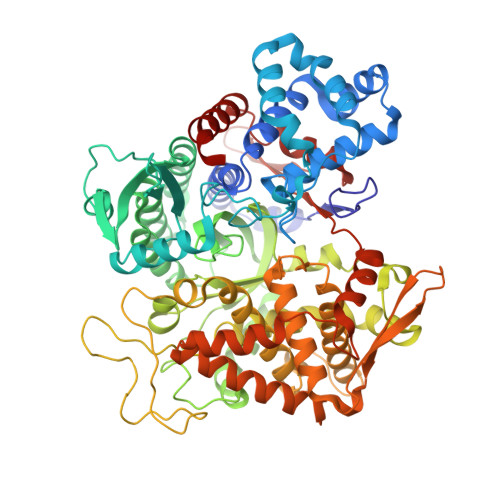
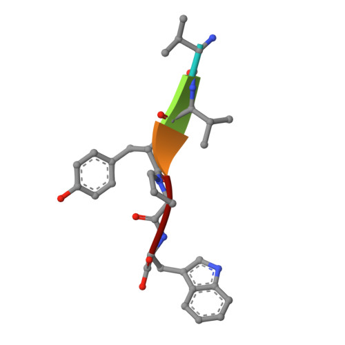
Entropy-driven binding of opioid peptides induces a large domain motion in human dipeptidyl peptidase III

Bezerra, G.A.Dobrovetsky, E.Viertlmayr, R.Dong, A.Binter, A.Abramic, M.Macheroux, P.Dhe-Paganon, S.Gruber, K.

(2012) Proc Natl Acad Sci U S A 109: 6525-6530

  • DOI: https://doi.org/10.1073/pnas.1118005109
  • Primary Citation of Related Structures:  
    3FVY, 3T6B, 3T6J

  • PubMed Abstract: 

    Opioid peptides are involved in various essential physiological processes, most notably nociception. Dipeptidyl peptidase III (DPP III) is one of the most important enkephalin-degrading enzymes associated with the mammalian pain modulatory system. Here we describe the X-ray structures of human DPP III and its complex with the opioid peptide tynorphin, which rationalize the enzyme's substrate specificity and reveal an exceptionally large domain motion upon ligand binding. Microcalorimetric analyses point at an entropy-dominated process, with the release of water molecules from the binding cleft ("entropy reservoir") as the major thermodynamic driving force. Our results provide the basis for the design of specific inhibitors that enable the elucidation of the exact role of DPP III and the exploration of its potential as a target of pain intervention strategies.


  • Organizational Affiliation

    Institute of Molecular Biosciences, University of Graz, A-8010 Graz, Austria.


Macromolecules
Find similar proteins by:  (by identity cutoff)  |  3D Structure
Entity ID: 1
MoleculeChains Sequence LengthOrganismDetailsImage
Dipeptidyl peptidase 3A,
C [auth B]
726Homo sapiensMutation(s): 1 
Gene Names: DPP3hDPPIII
EC: 3.4.14.4
UniProt & NIH Common Fund Data Resources
Find proteins for Q9NY33 (Homo sapiens)
Explore Q9NY33 
Go to UniProtKB:  Q9NY33
PHAROS:  Q9NY33
GTEx:  ENSG00000254986 
Entity Groups  
Sequence Clusters30% Identity50% Identity70% Identity90% Identity95% Identity100% Identity
UniProt GroupQ9NY33
Sequence Annotations
Expand
  • Reference Sequence

Find similar proteins by:  Sequence   |   3D Structure  

Entity ID: 2
MoleculeChains Sequence LengthOrganismDetailsImage
TynorphinB [auth C],
D
5Homo sapiensMutation(s): 0 
Sequence Annotations
Expand
  • Reference Sequence
Experimental Data & Validation

Experimental Data

  • Method: X-RAY DIFFRACTION
  • Resolution: 2.40 Å
  • R-Value Free: 0.258 
  • R-Value Work: 0.197 
  • R-Value Observed: 0.200 
  • Space Group: P 1 21 1
Unit Cell:
Length ( Å )Angle ( ˚ )
a = 61.97α = 90
b = 107.55β = 90.67
c = 119.1γ = 90
Software Package:
Software NamePurpose
MOSFLMdata reduction
SCALAdata scaling
PHASERphasing
PHENIXrefinement
PDB_EXTRACTdata extraction
DNAdata collection

Structure Validation

View Full Validation Report


Entry History 

Deposition Data

Revision History  (Full details and data files)

  • Version 1.0: 2012-04-04
    Type: Initial release
  • Version 1.1: 2012-04-18
    Changes: Database references
  • Version 1.2: 2012-12-12
    Changes: Other
  • Version 1.3: 2013-03-13
    Changes: Other
  • Version 1.4: 2013-06-19
    Changes: Database references
  • Version 1.5: 2023-11-01
    Changes: Data collection, Database references, Refinement description