3T5O

Crystal Structure of human Complement Component C6


Experimental Data Snapshot

  • Method: X-RAY DIFFRACTION
  • Resolution: 2.87 Å
  • R-Value Free: 0.276 
  • R-Value Work: 0.222 
  • R-Value Observed: 0.225 

wwPDB Validation   3D Report Full Report


Ligand Structure Quality Assessment 


This is version 2.0 of the entry. See complete history


Literature

Structure of Complement C6 Suggests a Mechanism for Initiation and Unidirectional, Sequential Assembly of Membrane Attack Complex (MAC).

Aleshin, A.E.Schraufstatter, I.U.Stec, B.Bankston, L.A.Liddington, R.C.Discipio, R.G.

(2012) J Biol Chem 287: 10210-10222

  • DOI: https://doi.org/10.1074/jbc.M111.327809
  • Primary Citation of Related Structures:  
    3T5O

  • PubMed Abstract: 

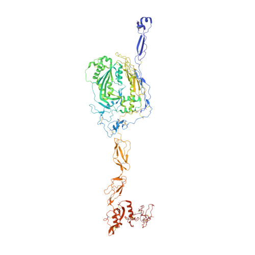
    The complement membrane attack complex (MAC) is formed by the sequential assembly of C5b with four homologous proteins as follows: one copy each of C6, C7, and C8 and 12-14 copies of C9. Together these form a lytic pore in bacterial membranes. C6 through C9 comprise a MAC-perforin domain flanked by 4-9 "auxiliary" domains. Here, we report the crystal structure of C6, the first and longest of the pore proteins to be recruited by C5b. Comparisons with the structures of the C8αβγ heterodimer and perforin show that the central domain of C6 adopts a "closed" (perforin-like) state that is distinct from the "open" conformations in C8. We further show that C6, C8α, and C8β contain three homologous subdomains ("upper," "lower," and "regulatory") related by rotations about two hinge points. In C6, the regulatory segment includes four auxiliary domains that stabilize the closed conformation, inhibiting release of membrane-inserting elements. In C8β, rotation of the regulatory segment is linked to an opening of the central β-sheet of its clockwise partner, C8α. Based on these observations, we propose a model for initiation and unidirectional propagation of the MAC in which the auxiliary domains play key roles: in the assembly of the C5b-8 initiation complex; in driving and regulating the opening of the β-sheet of the MAC-performin domain of each new recruit as it adds to the growing pore; and in stabilizing the final pore. Our model of the assembled pore resembles those of the cholesterol-dependent cytolysins but is distinct from that recently proposed for perforin.


  • Organizational Affiliation

    Infectious and Inflammatory Disease Center, Sanford-Burnham Medical Research Institute, La Jolla, California 92037 and.


Macromolecules
Find similar proteins by:  (by identity cutoff)  |  3D Structure
Entity ID: 1
MoleculeChains Sequence LengthOrganismDetailsImage
Complement component C6913Homo sapiensMutation(s): 0 
UniProt & NIH Common Fund Data Resources
Find proteins for P13671 (Homo sapiens)
Explore P13671 
Go to UniProtKB:  P13671
PHAROS:  P13671
GTEx:  ENSG00000039537 
Entity Groups  
Sequence Clusters30% Identity50% Identity70% Identity90% Identity95% Identity100% Identity
UniProt GroupP13671
Sequence Annotations
Expand
  • Reference Sequence
Oligosaccharides

Help

Entity ID: 2
MoleculeChains Length2D Diagram Glycosylation3D Interactions
2-acetamido-2-deoxy-beta-D-glucopyranose-(1-4)-2-acetamido-2-deoxy-beta-D-glucopyranose
B
2N-Glycosylation
Glycosylation Resources
GlyTouCan:  G42666HT
GlyCosmos:  G42666HT
GlyGen:  G42666HT
Entity ID: 3
MoleculeChains Length2D Diagram Glycosylation3D Interactions
beta-D-glucopyranose-(1-3)-alpha-L-fucopyranose
C
2O-Glycosylation
Glycosylation Resources
GlyTouCan:  G36855WW
GlyCosmos:  G36855WW
GlyGen:  G36855WW
Experimental Data & Validation

Experimental Data

  • Method: X-RAY DIFFRACTION
  • Resolution: 2.87 Å
  • R-Value Free: 0.276 
  • R-Value Work: 0.222 
  • R-Value Observed: 0.225 
  • Space Group: P 21 21 2
Unit Cell:
Length ( Å )Angle ( ˚ )
a = 146.81α = 90
b = 180.15β = 90
c = 60.53γ = 90
Software Package:
Software NamePurpose
Blu-Icedata collection
SHARPphasing
PHENIXrefinement
MOSFLMdata reduction
SCALAdata scaling

Structure Validation

View Full Validation Report


Ligand Structure Quality Assessment 


Entry History 

Deposition Data

Revision History  (Full details and data files)

  • Version 1.0: 2012-02-01
    Type: Initial release
  • Version 1.1: 2012-02-08
    Changes: Database references
  • Version 1.2: 2012-04-11
    Changes: Database references
  • Version 2.0: 2020-07-29
    Type: Remediation
    Reason: Carbohydrate remediation
    Changes: Advisory, Atomic model, Data collection, Derived calculations, Structure summary