3SZM

STRUCTURE OF HUMAN MICROCEPHALIN (MCPH1) TANDEM BRCT DOMAINS IN COMPLEX WITH A GAMMA-H2AX PHOSPHOPEPTIDE


Experimental Data Snapshot

  • Method: X-RAY DIFFRACTION
  • Resolution: 2.63 Å
  • R-Value Free: 0.252 
  • R-Value Work: 0.201 
  • R-Value Observed: 0.202 

wwPDB Validation   3D Report Full Report


This is version 1.2 of the entry. See complete history


Literature

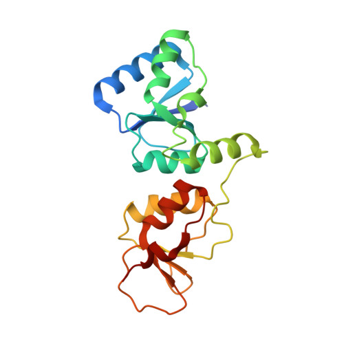
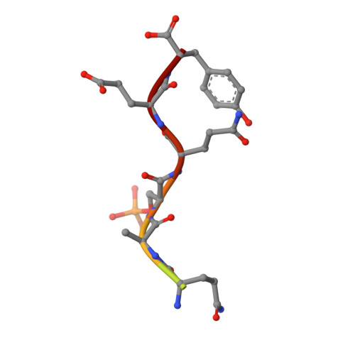
Dual recognition of phosphoserine and phosphotyrosine in histone variant H2A.X by DNA damage response protein MCPH1.

Singh, N.Basnet, H.Wiltshire, T.D.Mohammad, D.H.Thompson, J.R.Heroux, A.Botuyan, M.V.Yaffe, M.B.Couch, F.J.Rosenfeld, M.G.Mer, G.

(2012) Proc Natl Acad Sci U S A 109: 14381-14386

  • DOI: https://doi.org/10.1073/pnas.1212366109
  • Primary Citation of Related Structures:  
    3SZM, 3U3Z

  • PubMed Abstract: 

    Tyr142, the C-terminal amino acid of histone variant H2A.X is phosphorylated by WSTF (Williams-Beuren syndrome transcription factor), a component of the WICH complex (WSTF-ISWI chromatin-remodeling complex), under basal conditions in the cell. In response to DNA double-strand breaks (DSBs), H2A.X is instantaneously phosphorylated at Ser139 by the kinases ATM and ATR and is progressively dephosphorylated at Tyr142 by the Eya1 and Eya3 tyrosine phosphatases, resulting in a temporal switch from a postulated diphosphorylated (pSer139, pTyr142) to monophosphorylated (pSer139) H2A.X state. How mediator proteins interpret these two signals remains a question of fundamental interest. We provide structural, biochemical, and cellular evidence that Microcephalin (MCPH1), an early DNA damage response protein, can read both modifications via its tandem BRCA1 C-terminal (BRCT) domains, thereby emerging as a versatile sensor of H2A.X phosphorylation marks. We show that MCPH1 recruitment to sites of DNA damage is linked to both states of H2A.X.


  • Organizational Affiliation

    Department of Biochemistry and Molecular Biology, Mayo Clinic, Rochester, MN 55905, USA.


Macromolecules
Find similar proteins by:  (by identity cutoff)  |  3D Structure
Entity ID: 1
MoleculeChains Sequence LengthOrganismDetailsImage
Microcephalin
A, B, C, D, E
A, B, C, D, E, F, G, H
199Homo sapiensMutation(s): 0 
Gene Names: MCPH1
UniProt & NIH Common Fund Data Resources
Find proteins for Q8NEM0 (Homo sapiens)
Explore Q8NEM0 
Go to UniProtKB:  Q8NEM0
PHAROS:  Q8NEM0
GTEx:  ENSG00000147316 
Entity Groups  
Sequence Clusters30% Identity50% Identity70% Identity90% Identity95% Identity100% Identity
UniProt GroupQ8NEM0
Sequence Annotations
Expand
  • Reference Sequence

Find similar proteins by:  Sequence   |   3D Structure  

Entity ID: 2
MoleculeChains Sequence LengthOrganismDetailsImage
Histone H2A.x
I, J, K, L, M
I, J, K, L, M, N, O, P
10Homo sapiensMutation(s): 0 
UniProt & NIH Common Fund Data Resources
Find proteins for P16104 (Homo sapiens)
Explore P16104 
Go to UniProtKB:  P16104
PHAROS:  P16104
GTEx:  ENSG00000188486 
Entity Groups  
Sequence Clusters30% Identity50% Identity70% Identity90% Identity95% Identity100% Identity
UniProt GroupP16104
Sequence Annotations
Expand
  • Reference Sequence
Small Molecules
Modified Residues  1 Unique
IDChains TypeFormula2D DiagramParent
SEP
Query on SEP
I, J, K, L, M
I, J, K, L, M, N, O, P
L-PEPTIDE LINKINGC3 H8 N O6 PSER
Experimental Data & Validation

Experimental Data

  • Method: X-RAY DIFFRACTION
  • Resolution: 2.63 Å
  • R-Value Free: 0.252 
  • R-Value Work: 0.201 
  • R-Value Observed: 0.202 
  • Space Group: P 1
Unit Cell:
Length ( Å )Angle ( ˚ )
a = 67.81α = 72.1
b = 82.23β = 89.87
c = 104.42γ = 80.94
Software Package:
Software NamePurpose
HKL-2000data collection
DMmodel building
PHENIXrefinement
HKL-2000data reduction
HKL-2000data scaling
DMphasing

Structure Validation

View Full Validation Report


Entry History 

Deposition Data

Revision History  (Full details and data files)

  • Version 1.0: 2011-11-30
    Type: Initial release
  • Version 1.1: 2012-08-29
    Changes: Database references
  • Version 1.2: 2012-09-19
    Changes: Database references