3SX0

Crystal structure of Dot1l in complex with a brominated SAH analog


Experimental Data Snapshot

  • Method: X-RAY DIFFRACTION
  • Resolution: 2.28 Å
  • R-Value Free: 0.228 
  • R-Value Work: 0.209 
  • R-Value Observed: 0.210 

wwPDB Validation   3D Report Full Report


Ligand Structure Quality Assessment 


This is version 1.3 of the entry. See complete history


Literature

Bromo-deaza-SAH: a potent and selective DOT1L inhibitor.

Yu, W.Smil, D.Li, F.Tempel, W.Fedorov, O.Nguyen, K.T.Bolshan, Y.Al-Awar, R.Knapp, S.Arrowsmith, C.H.Vedadi, M.Brown, P.J.Schapira, M.

(2013) Bioorg Med Chem 21: 1787-1794

  • DOI: https://doi.org/10.1016/j.bmc.2013.01.049
  • Primary Citation of Related Structures:  
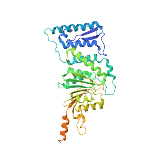
    3SX0

  • PubMed Abstract: 

    Chemical inhibition of proteins involved in chromatin-mediated signaling is an emerging strategy to control chromatin compaction with the aim to reprogram expression networks to alter disease states. Protein methyltransferases constitute one of the protein families that participate in epigenetic control of gene expression, and represent a novel therapeutic target class. Recruitment of the protein lysine methyltransferase DOT1L at aberrant loci is a frequent mechanism driving acute lymphoid and myeloid leukemias, particularly in infants, and pharmacological inhibition of DOT1L extends survival in a mouse model of mixed lineage leukemia. A better understanding of the structural chemistry of DOT1L inhibition would accelerate the development of improved compounds. Here, we report that the addition of a single halogen atom at a critical position in the cofactor product S-adenosylhomocysteine (SAH, an inhibitor of SAM-dependent methyltransferases) results in an 8-fold increase in potency against DOT1L, and reduced activities against other protein and non-protein methyltransferases. We solved the crystal structure of DOT1L in complex with Bromo-deaza-SAH and rationalized the observed effects. This discovery reveals a simple strategy to engineer selectivity and potency towards DOT1L into the adenosine scaffold of the cofactor shared by all methyltransferases, and can be exploited towards the development of clinical candidates against mixed lineage leukemia.


  • Organizational Affiliation

    Structural Genomics Consortium, University of Toronto; 101 College Street, MaRS Centre, South tower, Toronto, Ontario, M5G 1L7, Canada.


Macromolecules
Find similar proteins by:  (by identity cutoff)  |  3D Structure
Entity ID: 1
MoleculeChains Sequence LengthOrganismDetailsImage
Histone-lysine N-methyltransferase, H3 lysine-79 specific421Homo sapiensMutation(s): 0 
Gene Names: DOT1LKIAA1814KMT4
EC: 2.1.1.43
UniProt & NIH Common Fund Data Resources
Find proteins for Q8TEK3 (Homo sapiens)
Explore Q8TEK3 
Go to UniProtKB:  Q8TEK3
PHAROS:  Q8TEK3
GTEx:  ENSG00000104885 
Entity Groups  
Sequence Clusters30% Identity50% Identity70% Identity90% Identity95% Identity100% Identity
UniProt GroupQ8TEK3
Sequence Annotations
Expand
  • Reference Sequence
Small Molecules
Ligands 3 Unique
IDChains Name / Formula / InChI Key2D Diagram3D Interactions
SX0
Query on SX0

Download Ideal Coordinates CCD File 
B [auth A](2S)-2-amino-4-({[(2S,3S,4R,5R)-5-(4-amino-5-bromo-7H-pyrrolo[2,3-d]pyrimidin-7-yl)-3,4-dihydroxytetrahydrofuran-2-yl]methyl}sulfanyl)butanoic acid (non-preferred name)
C15 H20 Br N5 O5 S
DIULHULFPSIBAK-TWBCTODHSA-N
SO4
Query on SO4

Download Ideal Coordinates CCD File 
C [auth A]SULFATE ION
O4 S
QAOWNCQODCNURD-UHFFFAOYSA-L
UNX
Query on UNX

Download Ideal Coordinates CCD File 
D [auth A]
E [auth A]
F [auth A]
G [auth A]
H [auth A]
D [auth A],
E [auth A],
F [auth A],
G [auth A],
H [auth A],
I [auth A],
J [auth A],
K [auth A],
L [auth A],
M [auth A],
N [auth A],
O [auth A],
P [auth A],
Q [auth A],
R [auth A],
S [auth A],
T [auth A],
U [auth A],
V [auth A],
W [auth A]
UNKNOWN ATOM OR ION
X
Binding Affinity Annotations 
IDSourceBinding Affinity
SX0 BindingDB:  3SX0 Ki: 38 (nM) from 1 assay(s)
IC50: 77 (nM) from 1 assay(s)
Experimental Data & Validation

Experimental Data

  • Method: X-RAY DIFFRACTION
  • Resolution: 2.28 Å
  • R-Value Free: 0.228 
  • R-Value Work: 0.209 
  • R-Value Observed: 0.210 
  • Space Group: P 65
Unit Cell:
Length ( Å )Angle ( ˚ )
a = 152.917α = 90
b = 152.917β = 90
c = 50.964γ = 120
Software Package:
Software NamePurpose
DENZOdata reduction
SCALEPACKdata scaling
REFMACrefinement
PDB_EXTRACTdata extraction
HKL-2000data reduction
HKL-2000data scaling

Structure Validation

View Full Validation Report


Ligand Structure Quality Assessment 


Entry History 

Revision History  (Full details and data files)

  • Version 1.0: 2011-07-27
    Type: Initial release
  • Version 1.1: 2017-11-08
    Changes: Refinement description
  • Version 1.2: 2018-05-16
    Changes: Data collection, Database references
  • Version 1.3: 2023-09-13
    Changes: Data collection, Database references, Derived calculations, Refinement description