3SVJ

Strep Peptide Deformylase with a time dependent thiazolidine amide


Experimental Data Snapshot

  • Method: X-RAY DIFFRACTION
  • Resolution: 1.55 Å
  • R-Value Free: 0.208 
  • R-Value Work: 0.181 
  • R-Value Observed: 0.182 

wwPDB Validation   3D Report Full Report


Ligand Structure Quality Assessment 


This is version 1.3 of the entry. See complete history


Literature

Understanding the origins of time-dependent inhibition by polypeptide deformylase inhibitors.

Totoritis, R.Duraiswami, C.Taylor, A.N.Kerrigan, J.J.Campobasso, N.Smith, K.J.Ward, P.King, B.W.Murrayz-Thompson, M.Jones, A.D.Van Aller, G.S.Aubart, K.M.Zalacain, M.Thrall, S.H.Meek, T.D.Schwartz, B.

(2011) Biochemistry 50: 6642-6654

  • DOI: https://doi.org/10.1021/bi200655g
  • Primary Citation of Related Structures:  
    3STR, 3SVJ, 3SW8

  • PubMed Abstract: 

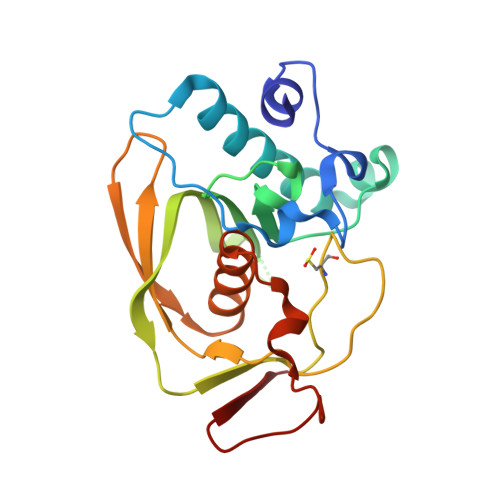
    The continual bacterial adaptation to antibiotics creates an ongoing medical need for the development of novel therapeutics. Polypeptide deformylase (PDF) is a highly conserved bacterial enzyme, which is essential for viability. It has previously been shown that PDF inhibitors represent a promising new area for the development of antimicrobial agents, and that many of the best PDF inhibitors demonstrate slow, time-dependent binding. To improve our understanding of the mechanistic origin of this time-dependent inhibition, we examined in detail the kinetics of PDF catalysis and inhibition by several different PDF inhibitors. Varying pH and solvent isotope led to clear changes in time-dependent inhibition parameters, as did inclusion of NaCl, which binds to the active site metal of PDF. Quantitative analysis of these results demonstrated that the observed time dependence arises from slow binding of the inhibitors to the active site metal. However, we also found several metal binding inhibitors that exhibited rapid, non-time-dependent onset of inhibition. By a combination of structural and chemical modification studies, we show that metal binding is only slow when the rest of the inhibitor makes optimal hydrogen bonds within the subsites of PDF. Both of these interactions between the inhibitor and enzyme were found to be necessary to observe time-dependent inhibition, as elimination of either leads to its loss.


  • Organizational Affiliation

    Department of Biological Reagents, GlaxoSmithKline, 1250 South Collegeville Road, Collegeville, Pennsylvania 19426, USA.


Macromolecules
Find similar proteins by:  (by identity cutoff)  |  3D Structure
Entity ID: 1
MoleculeChains Sequence LengthOrganismDetailsImage
Peptide deformylase 3A [auth P]203Streptococcus pneumoniaeMutation(s): 0 
Gene Names: defBdef3
EC: 3.5.1.88
UniProt
Find proteins for Q8DP79 (Streptococcus pneumoniae (strain ATCC BAA-255 / R6))
Explore Q8DP79 
Go to UniProtKB:  Q8DP79
Entity Groups  
Sequence Clusters30% Identity50% Identity70% Identity90% Identity95% Identity100% Identity
UniProt GroupQ8DP79
Sequence Annotations
Expand
  • Reference Sequence
Small Molecules
Ligands 4 Unique
IDChains Name / Formula / InChI Key2D Diagram3D Interactions
4LI
Query on 4LI

Download Ideal Coordinates CCD File 
E [auth P](4R)-3-(4-[4-(2-chlorophenyl)piperazin-1-yl]-6-{[2-methyl-6-(methylcarbamoyl)phenyl]amino}-1,3,5-triazin-2-yl)-N-methyl-1,3-thiazolidine-4-carboxamide
C27 H32 Cl N9 O2 S
QRGGVLXBPSQEDF-NRFANRHFSA-N
SO4
Query on SO4

Download Ideal Coordinates CCD File 
B [auth P],
C [auth P],
D [auth P]
SULFATE ION
O4 S
QAOWNCQODCNURD-UHFFFAOYSA-L
GOL
Query on GOL

Download Ideal Coordinates CCD File 
G [auth P]GLYCEROL
C3 H8 O3
PEDCQBHIVMGVHV-UHFFFAOYSA-N
NI
Query on NI

Download Ideal Coordinates CCD File 
F [auth P]NICKEL (II) ION
Ni
VEQPNABPJHWNSG-UHFFFAOYSA-N
Modified Residues  1 Unique
IDChains TypeFormula2D DiagramParent
OCS
Query on OCS
A [auth P]L-PEPTIDE LINKINGC3 H7 N O5 SCYS
Binding Affinity Annotations 
IDSourceBinding Affinity
4LI BindingDB:  3SVJ Ki: 117 (nM) from 1 assay(s)
Binding MOAD:  3SVJ Ki: 117 (nM) from 1 assay(s)
Experimental Data & Validation

Experimental Data

  • Method: X-RAY DIFFRACTION
  • Resolution: 1.55 Å
  • R-Value Free: 0.208 
  • R-Value Work: 0.181 
  • R-Value Observed: 0.182 
  • Space Group: P 43
Unit Cell:
Length ( Å )Angle ( ˚ )
a = 50.385α = 90
b = 50.385β = 90
c = 92.472γ = 90
Software Package:
Software NamePurpose
ADSCdata collection
PHASERphasing
PHENIXrefinement
HKL-2000data reduction
HKL-2000data scaling

Structure Validation

View Full Validation Report


Ligand Structure Quality Assessment 


Entry History 

Deposition Data

Revision History  (Full details and data files)

  • Version 1.0: 2011-07-27
    Type: Initial release
  • Version 1.1: 2011-08-17
    Changes: Database references
  • Version 1.2: 2014-04-16
    Changes: Other
  • Version 1.3: 2014-11-12
    Changes: Other