3SUN

RB69 DNA Polymerase (Y567A) Ternary Complex with dTTP Opposite 2AP (AT rich sequence)


Experimental Data Snapshot

  • Method: X-RAY DIFFRACTION
  • Resolution: 2.42 Å
  • R-Value Free: 0.255 
  • R-Value Work: 0.205 
  • R-Value Observed: 0.208 

wwPDB Validation   3D Report Full Report


Ligand Structure Quality Assessment 


This is version 1.3 of the entry. See complete history


Literature

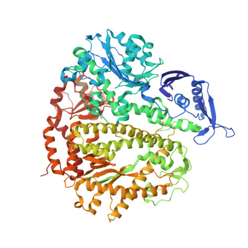
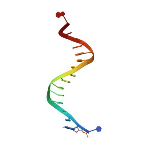
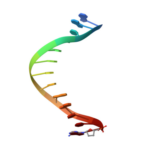
Structure of the 2-Aminopurine-Cytosine Base Pair Formed in the Polymerase Active Site of the RB69 Y567A-DNA Polymerase.

Reha-Krantz, L.J.Hariharan, C.Subuddhi, U.Xia, S.Zhao, C.Beckman, J.Christian, T.Konigsberg, W.

(2011) Biochemistry 50: 10136-10149

  • DOI: https://doi.org/10.1021/bi2014618
  • Primary Citation of Related Structures:  
    3SQ2, 3SQ4, 3SUN, 3SUO, 3SUP, 3SUQ

  • PubMed Abstract: 

    The adenine base analogue 2-aminopurine (2AP) is a potent base substitution mutagen in prokaryotes because of its enhanceed ability to form a mutagenic base pair with an incoming dCTP. Despite more than 50 years of research, the structure of the 2AP-C base pair remains unclear. We report the structure of the 2AP-dCTP base pair formed within the polymerase active site of the RB69 Y567A-DNA polymerase. A modified wobble 2AP-C base pair was detected with one H-bond between N1 of 2AP and a proton from the C4 amino group of cytosine and an apparent bifurcated H-bond between a proton on the 2-amino group of 2-aminopurine and the ring N3 and O2 atoms of cytosine. Interestingly, a primer-terminal region rich in AT base pairs, compared to GC base pairs, facilitated dCTP binding opposite template 2AP. We propose that the increased flexibility of the nucleotide binding pocket formed in the Y567A-DNA polymerase and increased "breathing" at the primer-terminal junction of A+T-rich DNA facilitate dCTP binding opposite template 2AP. Thus, interactions between DNA polymerase residues with a dynamic primer-terminal junction play a role in determining base selectivity within the polymerase active site of RB69 DNA polymerase.


  • Organizational Affiliation

    Department of Biological Sciences, University of Alberta, Edmonton, Alberta T6G 2E9, Canada. linda.reha-krantz@ualberta.ca


Macromolecules

Find similar proteins by:  (by identity cutoff)  |  3D Structure
Entity ID: 1
MoleculeChains Sequence LengthOrganismDetailsImage
DNA polymerase895Escherichia phage RB69Mutation(s): 3 
Gene Names: 43
EC: 2.7.7.7
UniProt
Find proteins for Q38087 (Escherichia phage RB69)
Explore Q38087 
Go to UniProtKB:  Q38087
Entity Groups  
Sequence Clusters30% Identity50% Identity70% Identity90% Identity95% Identity100% Identity
UniProt GroupQ38087
Sequence Annotations
Expand
  • Reference Sequence

Find similar nucleic acids by:  Sequence   |   3D Structure  

Entity ID: 2
MoleculeChains LengthOrganismImage
5'-D(P*CP*(2PR)P*TP*AP*AP*TP*TP*AP*AP*TP*TP*AP*AP*TP*TP*G)-3'B [auth T]16N/A
Sequence Annotations
Expand
  • Reference Sequence

Find similar nucleic acids by:  Sequence   |   3D Structure  

Entity ID: 3
MoleculeChains LengthOrganismImage
5'-D(*AP*AP*TP*TP*AP*AP*TP*TP*AP*AP*TP*TP*(2DA))-3'C [auth P]13N/A
Sequence Annotations
Expand
  • Reference Sequence
Experimental Data & Validation

Experimental Data

  • Method: X-RAY DIFFRACTION
  • Resolution: 2.42 Å
  • R-Value Free: 0.255 
  • R-Value Work: 0.205 
  • R-Value Observed: 0.208 
  • Space Group: P 21 21 21
Unit Cell:
Length ( Å )Angle ( ˚ )
a = 74.839α = 90
b = 120.987β = 90
c = 127.366γ = 90
Software Package:
Software NamePurpose
HKL-2000data collection
AMoREphasing
REFMACrefinement
HKL-2000data reduction
HKL-2000data scaling

Structure Validation

View Full Validation Report


Ligand Structure Quality Assessment 


Entry History 

Deposition Data

Revision History  (Full details and data files)

  • Version 1.0: 2011-11-09
    Type: Initial release
  • Version 1.1: 2011-11-16
    Changes: Database references
  • Version 1.2: 2011-11-30
    Changes: Database references
  • Version 1.3: 2024-02-28
    Changes: Data collection, Database references, Derived calculations