3SMZ

Human raver1 RRM1-3 domains (residues 39-320)


Experimental Data Snapshot

  • Method: X-RAY DIFFRACTION
  • Resolution: 1.99 Å
  • R-Value Free: 0.233 
  • R-Value Work: 0.185 
  • R-Value Observed: 0.188 

wwPDB Validation   3D Report Full Report


This is version 1.1 of the entry. See complete history


Literature

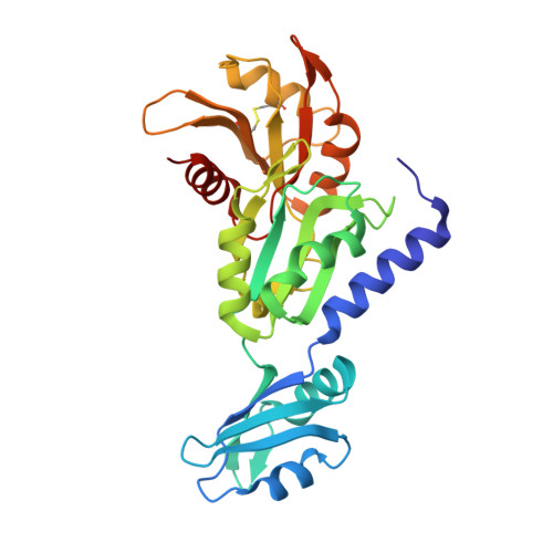
Apo raver1 structure reveals distinct RRM domain orientations.

Rangarajan, E.S.Lee, J.H.Izard, T.

(2011) Protein Sci 20: 1464-1470

  • DOI: https://doi.org/10.1002/pro.664
  • Primary Citation of Related Structures:  
    3SMZ

  • PubMed Abstract: 

    Raver1 is a multifunctional protein that modulates both alternative splicing and focal adhesion assembly by binding to the nucleoplasmic splicing repressor polypyrimidine tract protein (PTB) or to the cytoskeletal proteins vinculin and α-actinin. The amino-terminal region of raver1 has three RNA recognition motif (RRM1, RRM2, and RRM3) domains, and RRM1 interacts with the vinculin tail (Vt) domain and vinculin mRNA. We previously determined the crystal structure of the raver1 RRM1-3 domains in complex with Vt at 2.75 Å resolution. Here, we report crystal structure of the unbound raver1 RRM1-3 domains at 2 Å resolution. The apo structure reveals that a bound sulfate ion disrupts an electrostatic interaction between the RRM1 and RRM2 domains, triggering a large relative domain movement of over 30°. Superposition with other RNA-bound RRM structures places the sulfate ion near the superposed RNA phosphate group suggesting that this is the raver1 RNA binding site. While several single and some tandem RRM domain structures have been described, to the best of our knowledge, this is the second report of a three-tandem RRM domain structure.


  • Organizational Affiliation

    Cell Adhesion Laboratory, Department of Cancer Biology, The Scripps Research Institute, Jupiter, Florida 33458, USA.


Macromolecules
Find similar proteins by:  (by identity cutoff)  |  3D Structure
Entity ID: 1
MoleculeChains Sequence LengthOrganismDetailsImage
Ribonucleoprotein PTB-binding 1284Homo sapiensMutation(s): 0 
Gene Names: KIAA1978RAVER1
UniProt & NIH Common Fund Data Resources
Find proteins for Q8IY67 (Homo sapiens)
Explore Q8IY67 
Go to UniProtKB:  Q8IY67
PHAROS:  Q8IY67
Entity Groups  
Sequence Clusters30% Identity50% Identity70% Identity90% Identity95% Identity100% Identity
UniProt GroupQ8IY67
Sequence Annotations
Expand
  • Reference Sequence
Experimental Data & Validation

Experimental Data

  • Method: X-RAY DIFFRACTION
  • Resolution: 1.99 Å
  • R-Value Free: 0.233 
  • R-Value Work: 0.185 
  • R-Value Observed: 0.188 
  • Space Group: P 65
Unit Cell:
Length ( Å )Angle ( ˚ )
a = 104.02α = 90
b = 104.02β = 90
c = 50.42γ = 120
Software Package:
Software NamePurpose
SCALAdata scaling
PHASERphasing
BUSTER-TNTrefinement
PDB_EXTRACTdata extraction
SERGUIdata collection
XDSdata reduction
BUSTERrefinement

Structure Validation

View Full Validation Report


Entry History 

Deposition Data

Revision History  (Full details and data files)

  • Version 1.0: 2011-08-10
    Type: Initial release
  • Version 1.1: 2023-09-13
    Changes: Data collection, Database references, Derived calculations, Refinement description