3SMH

Crystal structure of major peanut allergen Ara h 1


Experimental Data Snapshot

  • Method: X-RAY DIFFRACTION
  • Resolution: 2.43 Å
  • R-Value Free: 0.244 
  • R-Value Work: 0.202 
  • R-Value Observed: 0.204 

wwPDB Validation   3D Report Full Report


This is version 1.1 of the entry. See complete history


Literature

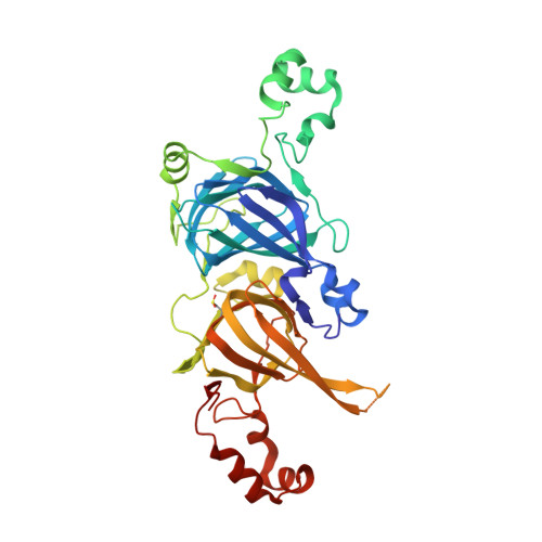
Crystal structure of the major peanut allergen Ara h 1.

Cabanos, C.Urabe, H.Tandang-Silvas, M.R.Utsumi, S.Mikami, B.Maruyama, N.

(2011) Mol Immunol 49: 115-123

  • DOI: https://doi.org/10.1016/j.molimm.2011.08.004
  • Primary Citation of Related Structures:  
    3SMH

  • PubMed Abstract: 

    Ara h 1, a 7S globulin, is one of the three major peanut allergens. We previously reported the crystallization of the core region of recombinant Ara h 1. Here, we present the crystal structure of the Ara h 1 core at a resolution of 2.43 Å. We also assayed the Ara h 1 core thermal stability and compared its final structure against other 7S globulins. The Ara h 1 core has a thermal denaturation temperature of 88.3°C and a structure that is very similar to other 7S globulins. Previously identified linear IgE epitopes were also mapped on the three-dimensional structure. Most linear epitopes were found in the extended loop domains and the coils between the N- and C-terminal modules, while others were found in the less accessible β-sheets of the C-terminal core β-barrel domain of each monomer. Most of these epitopes become either slightly or significantly buried upon trimer formation, implying that allergen digestion in the gut is required for these epitopes to be accessible to immunoglobulins. Our findings also suggest that both intact and partially degraded allergens should be employed in future diagnostic and immunotherapeutic strategies.


  • Organizational Affiliation

    Laboratory of Food Quality Design and Development, Graduate School of Agriculture, Kyoto University Uji, Kyoto, Japan.


Macromolecules
Find similar proteins by:  (by identity cutoff)  |  3D Structure
Entity ID: 1
MoleculeChains Sequence LengthOrganismDetailsImage
Allergen Ara h 1, clone P41B
A, B, C, D, E
A, B, C, D, E, F
418Arachis hypogaeaMutation(s): 0 
Gene Names: Arachis hypogea
UniProt
Find proteins for P43238 (Arachis hypogaea)
Explore P43238 
Go to UniProtKB:  P43238
Entity Groups  
Sequence Clusters30% Identity50% Identity70% Identity90% Identity95% Identity100% Identity
UniProt GroupP43238
Sequence Annotations
Expand
  • Reference Sequence
Small Molecules
Modified Residues  1 Unique
IDChains TypeFormula2D DiagramParent
CSD
Query on CSD
A, B, C, D, E
A, B, C, D, E, F
L-PEPTIDE LINKINGC3 H7 N O4 SCYS
Experimental Data & Validation

Experimental Data

  • Method: X-RAY DIFFRACTION
  • Resolution: 2.43 Å
  • R-Value Free: 0.244 
  • R-Value Work: 0.202 
  • R-Value Observed: 0.204 
  • Space Group: C 1 2 1
Unit Cell:
Length ( Å )Angle ( ˚ )
a = 156.492α = 90
b = 89.008β = 107.15
c = 158.966γ = 90
Software Package:
Software NamePurpose
HKL-2000data collection
CCP4model building
PHENIXrefinement
DENZOdata reduction
SCALEPACKdata scaling
CCP4phasing

Structure Validation

View Full Validation Report


Entry History 

Deposition Data

Revision History  (Full details and data files)

  • Version 1.0: 2012-02-15
    Type: Initial release
  • Version 1.1: 2014-11-12
    Changes: Non-polymer description