3SLK

Structure of ketoreductase and enoylreductase didomain from modular polyketide synthase


Experimental Data Snapshot

  • Method: X-RAY DIFFRACTION
  • Resolution: 3.00 Å
  • R-Value Free: 0.256 
  • R-Value Work: 0.210 
  • R-Value Observed: 0.212 

wwPDB Validation   3D Report Full Report


Ligand Structure Quality Assessment 


This is version 1.3 of the entry. See complete history


Literature

Divergence of multimodular polyketide synthases revealed by a didomain structure.

Zheng, J.Gay, D.C.Demeler, B.White, M.A.Keatinge-Clay, A.T.

(2012) Nat Chem Biol 8: 615-621

  • DOI: https://doi.org/10.1038/nchembio.964
  • Primary Citation of Related Structures:  
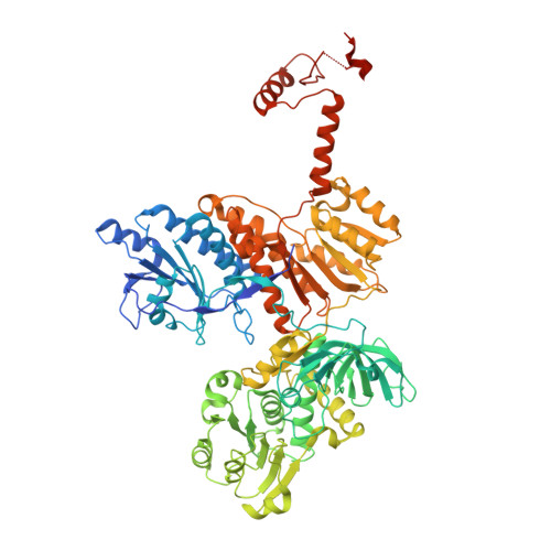
    3SLK

  • PubMed Abstract: 

    The enoylreductase (ER) is the final common enzyme from modular polyketide synthases (PKSs) to be structurally characterized. The 3.0 Å-resolution structure of the didomain comprising the ketoreductase (KR) and ER from the second module of the spinosyn PKS reveals that ER shares an ∼600-Å(2) interface with KR distinct from that of the related mammalian fatty acid synthase (FAS). In contrast to the ER domains of the mammalian FAS, the ER domains of the second module of the spinosyn PKS do not make contact across the two-fold axis of the synthase. This monomeric organization may have been necessary in the evolution of multimodular PKSs to enable acyl carrier proteins to access each of their cognate enzymes. The isolated ER domain showed activity toward a substrate analog, enabling us to determine the contributions of its active site residues.


  • Organizational Affiliation

    Department of Chemistry and Biochemistry, The University of Texas at Austin, Austin, Texas, USA.


Macromolecules
Find similar proteins by:  (by identity cutoff)  |  3D Structure
Entity ID: 1
MoleculeChains Sequence LengthOrganismDetailsImage
Polyketide synthase extender module 2
A, B
795Saccharopolyspora spinosaMutation(s): 0 
Gene Names: spnB
EC: 1.1.1.100 (PDB Primary Data), 1.3.1.9 (PDB Primary Data)
UniProt
Find proteins for Q9ALM5 (Saccharopolyspora spinosa)
Explore Q9ALM5 
Go to UniProtKB:  Q9ALM5
Entity Groups  
Sequence Clusters30% Identity50% Identity70% Identity90% Identity95% Identity100% Identity
UniProt GroupQ9ALM5
Sequence Annotations
Expand
  • Reference Sequence
Experimental Data & Validation

Experimental Data

  • Method: X-RAY DIFFRACTION
  • Resolution: 3.00 Å
  • R-Value Free: 0.256 
  • R-Value Work: 0.210 
  • R-Value Observed: 0.212 
  • Space Group: P 21 21 21
Unit Cell:
Length ( Å )Angle ( ˚ )
a = 73.104α = 90
b = 110.195β = 90
c = 202.857γ = 90
Software Package:
Software NamePurpose
HKL-2000data collection
SOLVEphasing
REFMACrefinement
HKL-2000data reduction
HKL-2000data scaling

Structure Validation

View Full Validation Report


Ligand Structure Quality Assessment 


Entry History 

Deposition Data

Revision History  (Full details and data files)

  • Version 1.0: 2012-05-30
    Type: Initial release
  • Version 1.1: 2012-06-13
    Changes: Database references
  • Version 1.2: 2012-09-05
    Changes: Database references
  • Version 1.3: 2024-02-28
    Changes: Data collection, Database references, Derived calculations