3SK2

Crystal structure of phenazine resistance protein EhpR from Enterobacter agglomerans (Erwinia herbicola, Pantoea agglomerans) Eh1087 in complex with griseoluteic acid


Experimental Data Snapshot

  • Method: X-RAY DIFFRACTION
  • Resolution: 1.01 Å
  • R-Value Free: 0.146 
  • R-Value Work: 0.126 
  • R-Value Observed: 0.127 

wwPDB Validation   3D Report Full Report


Ligand Structure Quality Assessment 


This is version 1.2 of the entry. See complete history


Literature

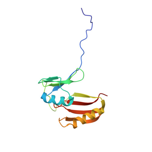
Atomic resolution structure of EhpR: phenazine resistance in Enterobacter agglomerans Eh1087 follows principles of bleomycin / mitomycin C resistance in other bacteria.

Yu, S.Vit, A.Devenish, S.Mahanty, H.K.Itzen, A.Goody, R.S.Blankenfeldt, W.

(2011) BMC Struct Biol 11: 33-33

  • DOI: https://doi.org/10.1186/1472-6807-11-33
  • Primary Citation of Related Structures:  
    3SK1, 3SK2

  • PubMed Abstract: 

    The phenazines are redox-active secondary metabolites that a large number of bacterial strains produce and excrete into the environment. They possess antibiotic activity owing to the fact that they can reduce molecular oxygen to toxic reactive oxygen species. In order to take advantage of this activity, phenazine producers need to protect themselves against phenazine toxicity. Whereas it is believed that phenazine-producing pseudomonads possess highly active superoxide dismutases and catalases, it has recently been found that the plant-colonizing bacterium Enterobacter agglomerans expresses a small gene ehpR to render itself resistant towards D-alanyl-griseoluteic acid, the phenazine antibiotic produced by this strain.


  • Organizational Affiliation

    Department of Physical Biochemistry, Max Planck Institute of Molecular Physiology, Otto-Hahn-Straße 11, 44227 Dortmund, Germany.


Macromolecules
Find similar proteins by:  (by identity cutoff)  |  3D Structure
Entity ID: 1
MoleculeChains Sequence LengthOrganismDetailsImage
EhpR
A, B
132Pantoea agglomeransMutation(s): 0 
Gene Names: ehpR
UniProt
Find proteins for Q8GPH6 (Enterobacter agglomerans)
Explore Q8GPH6 
Go to UniProtKB:  Q8GPH6
Entity Groups  
Sequence Clusters30% Identity50% Identity70% Identity90% Identity95% Identity100% Identity
UniProt GroupQ8GPH6
Sequence Annotations
Expand
  • Reference Sequence
Small Molecules
Ligands 1 Unique
IDChains Name / Formula / InChI Key2D Diagram3D Interactions
GRI
Query on GRI

Download Ideal Coordinates CCD File 
C [auth A]6-formyl-9-methoxyphenazine-1-carboxylic acid
C15 H10 N2 O4
NJJLLHHIAVHAEL-UHFFFAOYSA-N
Binding Affinity Annotations 
IDSourceBinding Affinity
GRI Binding MOAD:  3SK2 Kd: 2.44e+5 (nM) from 1 assay(s)
PDBBind:  3SK2 Kd: 2.44e+5 (nM) from 1 assay(s)
Experimental Data & Validation

Experimental Data

  • Method: X-RAY DIFFRACTION
  • Resolution: 1.01 Å
  • R-Value Free: 0.146 
  • R-Value Work: 0.126 
  • R-Value Observed: 0.127 
  • Space Group: P 21 21 21
Unit Cell:
Length ( Å )Angle ( ˚ )
a = 36.5α = 90
b = 79.77β = 90
c = 82.85γ = 90
Software Package:
Software NamePurpose
XDSdata scaling
PHASERphasing
PHENIXrefinement
XDSdata reduction
XSCALEdata scaling

Structure Validation

View Full Validation Report


Ligand Structure Quality Assessment 


Entry History 

Deposition Data

Revision History  (Full details and data files)

  • Version 1.0: 2011-08-31
    Type: Initial release
  • Version 1.1: 2024-02-28
    Changes: Data collection, Database references, Derived calculations
  • Version 1.2: 2024-04-03
    Changes: Refinement description