3SIQ

Crystal Structure of autoinhibited dIAP1-BIR1 domain


Experimental Data Snapshot

  • Method: X-RAY DIFFRACTION
  • Resolution: 2.40 Å
  • R-Value Free: 0.217 
  • R-Value Work: 0.146 
  • R-Value Observed: 0.150 

wwPDB Validation   3D Report Full Report


This is version 1.3 of the entry. See complete history


Literature

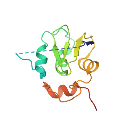
Structural mechanisms of DIAP1 auto-inhibition and DIAP1-mediated inhibition of drICE.

Li, X.Wang, J.Shi, Y.

(2011) Nat Commun 2: 408-408

  • DOI: https://doi.org/10.1038/ncomms1418
  • Primary Citation of Related Structures:  
    3SIP, 3SIQ, 3SIR

  • PubMed Abstract: 

    The Drosophila inhibitor of apoptosis protein DIAP1 exists in an auto-inhibited conformation, unable to suppress the effector caspase drICE. Auto-inhibition is disabled by caspase-mediated cleavage of DIAP1 after Asp20. The cleaved DIAP1 binds to mature drICE, inhibits its protease activity, and, presumably, also targets drICE for ubiquitylation. DIAP1-mediated suppression of drICE is effectively antagonized by the pro-apoptotic proteins Reaper, Hid, and Grim (RHG). Despite rigorous effort, the molecular mechanisms behind these observations are enigmatic. Here we report a 2.4 Å crystal structure of uncleaved DIAP1-BIR1, which reveals how the amino-terminal sequences recognize a conserved surface groove in BIR1 to achieve auto-inhibition, and a 3.5 Å crystal structure of active drICE bound to cleaved DIAP1-BIR1, which provides a structural explanation to DIAP1-mediated inhibition of drICE. These structures and associated biochemical analyses, together with published reports, define the molecular determinants that govern the interplay among DIAP1, drICE and the RHG proteins.


  • Organizational Affiliation

    Ministry of Education Protein Science Laboratory, Center for Structural Biology, School of Life Sciences and School of Medicine, Tsinghua University, Beijing, China.


Macromolecules
Find similar proteins by:  (by identity cutoff)  |  3D Structure
Entity ID: 1
MoleculeChains Sequence LengthOrganismDetailsImage
Apoptosis 1 inhibitor
A, B, C, D, E
A, B, C, D, E, F
136Drosophila melanogasterMutation(s): 1 
EC: 6.3.2
UniProt
Find proteins for Q24306 (Drosophila melanogaster)
Explore Q24306 
Go to UniProtKB:  Q24306
Entity Groups  
Sequence Clusters30% Identity50% Identity70% Identity90% Identity95% Identity100% Identity
UniProt GroupQ24306
Sequence Annotations
Expand
  • Reference Sequence
Experimental Data & Validation

Experimental Data

  • Method: X-RAY DIFFRACTION
  • Resolution: 2.40 Å
  • R-Value Free: 0.217 
  • R-Value Work: 0.146 
  • R-Value Observed: 0.150 
  • Space Group: P 31
Unit Cell:
Length ( Å )Angle ( ˚ )
a = 99.782α = 90
b = 99.782β = 90
c = 71.344γ = 120
Software Package:
Software NamePurpose
REFMACrefinement

Structure Validation

View Full Validation Report


Entry History 

Deposition Data

Revision History  (Full details and data files)

  • Version 1.0: 2011-08-10
    Type: Initial release
  • Version 1.1: 2013-07-10
    Changes: Database references
  • Version 1.2: 2018-07-25
    Changes: Data collection, Source and taxonomy, Structure summary
  • Version 1.3: 2023-11-01
    Changes: Data collection, Database references, Derived calculations, Refinement description