3S7M

Pyrazolyl and Thienyl Aminohydantoins as Potent BACE1 Inhibitors


Experimental Data Snapshot

  • Method: X-RAY DIFFRACTION
  • Resolution: 2.20 Å
  • R-Value Free: 0.245 
  • R-Value Work: 0.182 
  • R-Value Observed: 0.186 

wwPDB Validation   3D Report Full Report


Ligand Structure Quality Assessment 


This is version 1.0 of the entry. See complete history


Literature

New pyrazolyl and thienyl aminohydantoins as potent BACE1 inhibitors: Exploring the S2' region.

Malamas, M.S.Erdei, J.Gunawan, I.Barnes, K.Hui, Y.Johnson, M.Robichaud, A.Zhou, P.Yan, Y.Solvibile, W.Turner, J.Fan, K.Y.Chopra, R.Bard, J.Pangalos, M.N.

(2011) Bioorg Med Chem Lett 21: 5164-5170

  • DOI: https://doi.org/10.1016/j.bmcl.2011.07.057
  • Primary Citation of Related Structures:  
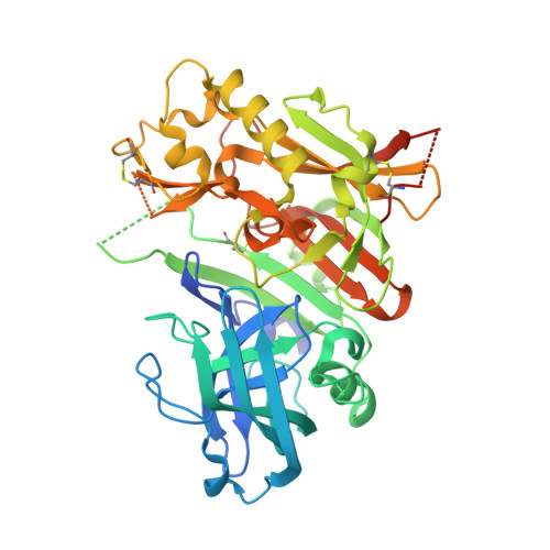
    3S7L, 3S7M

  • PubMed Abstract: 

    The proteolytic enzyme β-secretase (BACE1) plays a central role in the synthesis of the pathogenic β-amyloid in Alzheimer's disease. SAR studies of the S2' region of the BACE1 ligand binding pocket with pyrazolyl and thienyl P2' side chains are reported. These analogs exhibit low nanomolar potency for BACE1, and demonstrate >50- to 100-fold selectivity for the structurally related aspartyl proteases BACE2 and cathepsin D. Small groups attached at the nitrogen of the P2' pyrazolyl moiety, together with the P3 pyrimidine nucleus projecting into the S3 region of the binding pocket, are critical components to ligand's potency and selectivity. P2' thiophene side chain analogs are highly potent BACE1 inhibitors with excellent selectivity against cathepsin D, but only modest selectivity against BACE2. The cell-based activity of these new analogs tracked well with their increased molecular binding with EC(50) values of 0.07-0.2 μM in the ELISA assay for the most potent analogs.


  • Organizational Affiliation

    Pfizer Neuroscience Princeton, 865 Ridge Road, Monmouth Junction, NJ 08852, USA. malamas.michael@gmail.com


Macromolecules
Find similar proteins by:  (by identity cutoff)  |  3D Structure
Entity ID: 1
MoleculeChains Sequence LengthOrganismDetailsImage
Beta-secretase 1415Homo sapiensMutation(s): 0 
Gene Names: BACE1BACEKIAA1149
EC: 3.4.23.46
UniProt & NIH Common Fund Data Resources
Find proteins for P56817 (Homo sapiens)
Explore P56817 
Go to UniProtKB:  P56817
PHAROS:  P56817
GTEx:  ENSG00000186318 
Entity Groups  
Sequence Clusters30% Identity50% Identity70% Identity90% Identity95% Identity100% Identity
UniProt GroupP56817
Sequence Annotations
Expand
  • Reference Sequence
Small Molecules
Ligands 1 Unique
IDChains Name / Formula / InChI Key2D Diagram3D Interactions
532
Query on 532

Download Ideal Coordinates CCD File 
B [auth A](5S)-2-amino-3-methyl-5-[3-(pyridin-3-yl)phenyl]-5-(thiophen-3-yl)-3,5-dihydro-4H-imidazol-4-one
C19 H16 N4 O S
AOGBXAFIKRFUPJ-IBGZPJMESA-N
Binding Affinity Annotations 
IDSourceBinding Affinity
532 PDBBind:  3S7M IC50: 10 (nM) from 1 assay(s)
Binding MOAD:  3S7M IC50: 60 (nM) from 1 assay(s)
BindingDB:  3S7M IC50: 60 (nM) from 1 assay(s)
EC50: 420 (nM) from 1 assay(s)
Experimental Data & Validation

Experimental Data

  • Method: X-RAY DIFFRACTION
  • Resolution: 2.20 Å
  • R-Value Free: 0.245 
  • R-Value Work: 0.182 
  • R-Value Observed: 0.186 
  • Space Group: C 1 2 1
Unit Cell:
Length ( Å )Angle ( ˚ )
a = 72.389α = 90
b = 103.979β = 94.9
c = 50.137γ = 90
Software Package:
Software NamePurpose
CrystalCleardata collection
PHENIXmodel building
PHENIXrefinement
HKL-2000data reduction
HKL-2000data scaling
PHENIXphasing

Structure Validation

View Full Validation Report


Ligand Structure Quality Assessment 


Entry History 

Deposition Data

Revision History  (Full details and data files)

  • Version 1.0: 2011-08-31
    Type: Initial release