3S51

Structure of FANCI


Experimental Data Snapshot

  • Method: X-RAY DIFFRACTION
  • Resolution: 3.30 Å
  • R-Value Free: 0.278 
  • R-Value Work: 0.259 
  • R-Value Observed: 0.259 

wwPDB Validation   3D Report Full Report


This is version 1.2 of the entry. See complete history


Literature

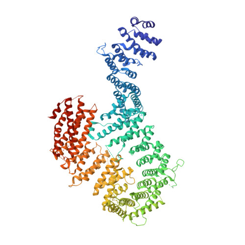
Structure of the FANCI-FANCD2 complex: insights into the Fanconi anemia DNA repair pathway.

Joo, W.Xu, G.Persky, N.S.Smogorzewska, A.Rudge, D.G.Buzovetsky, O.Elledge, S.J.Pavletich, N.P.

(2011) Science 333: 312-316

  • DOI: https://doi.org/10.1126/science.1205805
  • Primary Citation of Related Structures:  
    3S4W, 3S4Z, 3S51

  • PubMed Abstract: 

    Fanconi anemia is a cancer predisposition syndrome caused by defects in the repair of DNA interstrand cross-links (ICLs). Central to this pathway is the Fanconi anemia I-Fanconi anemia D2 (FANCI-FANCD2) (ID) complex, which is activated by DNA damage-induced phosphorylation and monoubiquitination. The 3.4 angstrom crystal structure of the ~300 kilodalton ID complex reveals that monoubiquitination and regulatory phosphorylation sites map to the I-D interface, suggesting that they occur on monomeric proteins or an opened-up complex and that they may serve to stabilize I-D heterodimerization. The 7.8 angstrom electron-density map of FANCI-DNA crystals and in vitro data show that each protein has binding sites for both single- and double-stranded DNA, suggesting that the ID complex recognizes DNA structures that result from the encounter of replication forks with an ICL.


  • Organizational Affiliation

    Structural Biology Program, Memorial Sloan-Kettering Cancer Center, New York, NY 10065, USA.


Macromolecules
Find similar proteins by:  (by identity cutoff)  |  3D Structure
Entity ID: 1
MoleculeChains Sequence LengthOrganismDetailsImage
Fanconi anemia group I protein homolog
A, B, C, D
1,308Mus musculusMutation(s): 0 
Gene Names: Fanci
UniProt & NIH Common Fund Data Resources
Find proteins for Q8K368 (Mus musculus)
Explore Q8K368 
Go to UniProtKB:  Q8K368
IMPC:  MGI:2384790
Entity Groups  
Sequence Clusters30% Identity50% Identity70% Identity90% Identity95% Identity100% Identity
UniProt GroupQ8K368
Sequence Annotations
Expand
  • Reference Sequence
Experimental Data & Validation

Experimental Data

  • Method: X-RAY DIFFRACTION
  • Resolution: 3.30 Å
  • R-Value Free: 0.278 
  • R-Value Work: 0.259 
  • R-Value Observed: 0.259 
  • Space Group: P 1
Unit Cell:
Length ( Å )Angle ( ˚ )
a = 114.7α = 115.9
b = 136.5β = 106
c = 149.7γ = 95
Software Package:
Software NamePurpose
HKL-2000data collection
SHARPphasing
PHENIXrefinement
HKL-2000data reduction
DPSdata reduction

Structure Validation

View Full Validation Report


Entry History 

Deposition Data

Revision History  (Full details and data files)

  • Version 1.0: 2011-07-27
    Type: Initial release
  • Version 1.1: 2012-03-28
    Changes: Database references
  • Version 1.2: 2024-02-28
    Changes: Data collection, Database references