3S4X

Crystal structure of the Asn152Gly mutant of P99 beta-lactamase


Experimental Data Snapshot

  • Method: X-RAY DIFFRACTION
  • Resolution: 1.95 Å
  • R-Value Free: 0.234 
  • R-Value Work: 0.176 
  • R-Value Observed: 0.178 

wwPDB Validation   3D Report Full Report


This is version 1.3 of the entry. See complete history


Literature

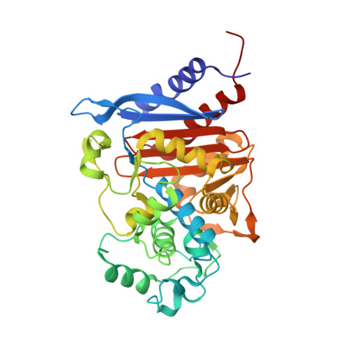
Structural analysis of the Asn152Gly mutant of P99 cephalosporinase.

Ruble, J.F.Lefurgy, S.T.Cornish, V.W.Powers, R.A.

(2012) Acta Crystallogr D Biol Crystallogr 68: 1189-1193

  • DOI: https://doi.org/10.1107/S0907444912024080
  • Primary Citation of Related Structures:  
    3S4X

  • PubMed Abstract: 

    P99 cephalosporinase is a class C β-lactamase that is responsible in part for the widespread bacterial resistance to β-lactam antibiotics. Mutations of the conserved active-site residue Asn152 of the enzyme have been shown to alter β-lactam substrate specificity in vivo. Mutation of Asn152 to a glycine is notable in that it exhibits in vivo substrate-selectivity switching. In order to better understand the structural basis for this observed switch, the X-ray crystal structure of the apo Asn152Gly mutant of P99 was determined to 1.95 Å resolution. Unexpectedly, the artificial C-terminal His(6) tag of a symmetrically-related molecule was observed bound in the active site. The His(6) tag makes several interactions with key active-site residues, as well as with several sulfate ions. Additionally, the overall C-terminus occupies the space left vacant upon the mutation of Asn152 to glycine.


  • Organizational Affiliation

    Department of Chemistry, Grand Valley State University, Allendale, MI 49401, USA.


Macromolecules
Find similar proteins by:  (by identity cutoff)  |  3D Structure
Entity ID: 1
MoleculeChains Sequence LengthOrganismDetailsImage
Beta-lactamase367Enterobacter cloacaeMutation(s): 4 
Gene Names: ampC
EC: 3.5.2.6
UniProt
Find proteins for P05364 (Enterobacter cloacae)
Explore P05364 
Go to UniProtKB:  P05364
Entity Groups  
Sequence Clusters30% Identity50% Identity70% Identity90% Identity95% Identity100% Identity
UniProt GroupP05364
Sequence Annotations
Expand
  • Reference Sequence
Experimental Data & Validation

Experimental Data

  • Method: X-RAY DIFFRACTION
  • Resolution: 1.95 Å
  • R-Value Free: 0.234 
  • R-Value Work: 0.176 
  • R-Value Observed: 0.178 
  • Space Group: P 21 21 2
Unit Cell:
Length ( Å )Angle ( ˚ )
a = 78.956α = 90
b = 85.481β = 90
c = 51.681γ = 90
Software Package:
Software NamePurpose
HKL-2000data collection
PHASERphasing
REFMACrefinement
HKL-2000data reduction
HKL-2000data scaling

Structure Validation

View Full Validation Report


Entry History 

Deposition Data

Revision History  (Full details and data files)

  • Version 1.0: 2012-08-01
    Type: Initial release
  • Version 1.1: 2012-08-29
    Changes: Database references
  • Version 1.2: 2013-01-23
    Changes: Database references
  • Version 1.3: 2023-09-13
    Changes: Data collection, Database references, Derived calculations, Refinement description