3S2U

Crystal structure of the Pseudomonas aeruginosa MurG:UDP-GlcNAc substrate complex


Experimental Data Snapshot

  • Method: X-RAY DIFFRACTION
  • Resolution: 2.23 Å
  • R-Value Free: 0.278 
  • R-Value Work: 0.209 
  • R-Value Observed: 0.214 

wwPDB Validation   3D Report Full Report


Ligand Structure Quality Assessment 


This is version 1.3 of the entry. See complete history


Literature

Crystal Structure of the Pseudomonas aeruginosa MurG: UDP-GlcNAc Substrate Complex.

Brown, K.Vial, S.C.Dedi, N.Westcott, J.Scally, S.Bugg, T.D.Charlton, P.A.Cheetham, G.M.

(2013) Protein Pept Lett 20: 1002-1008

  • DOI: https://doi.org/10.2174/0929866511320090006
  • Primary Citation of Related Structures:  
    3S2U

  • PubMed Abstract: 

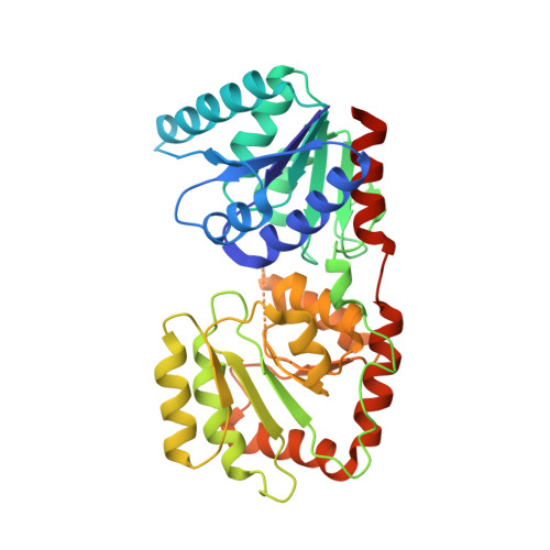
    MurG is an essential bacterial glycosyltransferase enzyme in Pseudomonas aeruginosa performing one of the key membrane steps of peptidoglycan synthesis catalyzing the transfer of N-acetyl glucosamine (GlcNAc) from its donor substrate, UDP-GlcNAc, to the acceptor substrate Lipid I. We have solved the crystal structure of the complex between Pseudomonas aeruginosa MurG and UDP-GlcNAc and compared it with the previously solved complex from E. coli. The structure reveals a large-scale conformational change in the relative orientations of the N- and C-terminal domains, which has the effect of widening the cofactor binding site and displacing the UDP-GlcNAc donor. These results suggest new opportunities to design potent inhibitors of peptidoglycan biosynthesis.


  • Organizational Affiliation

    Vertex Pharmaceuticals (Europe) Ltd, 88 Milton Park, Abingdon, Oxfordshire, OX14 4RY, UK. kieron_brown@vrtx.com


Macromolecules
Find similar proteins by:  (by identity cutoff)  |  3D Structure
Entity ID: 1
MoleculeChains Sequence LengthOrganismDetailsImage
UDP-N-acetylglucosamine--N-acetylmuramyl-(pentapeptide) pyrophosphoryl-undecaprenol N-acetylglucosamine transferase365Pseudomonas aeruginosa PAO1Mutation(s): 0 
Gene Names: murGPA4412
EC: 2.4.1.227
Membrane Entity: Yes 
UniProt
Find proteins for Q9HW01 (Pseudomonas aeruginosa (strain ATCC 15692 / DSM 22644 / CIP 104116 / JCM 14847 / LMG 12228 / 1C / PRS 101 / PAO1))
Explore Q9HW01 
Go to UniProtKB:  Q9HW01
Entity Groups  
Sequence Clusters30% Identity50% Identity70% Identity90% Identity95% Identity100% Identity
UniProt GroupQ9HW01
Sequence Annotations
Expand
  • Reference Sequence
Small Molecules
Ligands 1 Unique
IDChains Name / Formula / InChI Key2D Diagram3D Interactions
UD1
Query on UD1

Download Ideal Coordinates CCD File 
B [auth A]URIDINE-DIPHOSPHATE-N-ACETYLGLUCOSAMINE
C17 H27 N3 O17 P2
LFTYTUAZOPRMMI-CFRASDGPSA-N
Experimental Data & Validation

Experimental Data

  • Method: X-RAY DIFFRACTION
  • Resolution: 2.23 Å
  • R-Value Free: 0.278 
  • R-Value Work: 0.209 
  • R-Value Observed: 0.214 
  • Space Group: P 41 21 2
Unit Cell:
Length ( Å )Angle ( ˚ )
a = 49.93α = 90
b = 49.93β = 90
c = 278.62γ = 90
Software Package:
Software NamePurpose
DNAdata collection
AMoREphasing
CNSrefinement
MOSFLMdata reduction
SCALAdata scaling

Structure Validation

View Full Validation Report


Ligand Structure Quality Assessment 


Entry History 

Deposition Data

Revision History  (Full details and data files)

  • Version 1.0: 2012-09-26
    Type: Initial release
  • Version 1.1: 2013-05-08
    Changes: Database references
  • Version 1.2: 2013-08-14
    Changes: Database references
  • Version 1.3: 2024-02-28
    Changes: Data collection, Database references, Derived calculations