3RZI

The structure of 3-deoxy-D-arabino-heptulosonate 7-phosphate synthase from mycobacterium tuberculosis cocrystallized and complexed with phenylalanine and tryptophan


Experimental Data Snapshot

  • Method: X-RAY DIFFRACTION
  • Resolution: 1.95 Å
  • R-Value Free: 0.168 
  • R-Value Work: 0.153 
  • R-Value Observed: 0.154 

wwPDB Validation   3D Report Full Report


Ligand Structure Quality Assessment 


This is version 1.2 of the entry. See complete history


Literature

Dynamic cross-talk among remote binding sites: the molecular basis for unusual synergistic allostery.

Jiao, W.Hutton, R.D.Cross, P.J.Jameson, G.B.Parker, E.J.

(2012) J Mol Biol 415: 716-726

  • DOI: https://doi.org/10.1016/j.jmb.2011.11.037
  • Primary Citation of Related Structures:  
    3RZI

  • PubMed Abstract: 

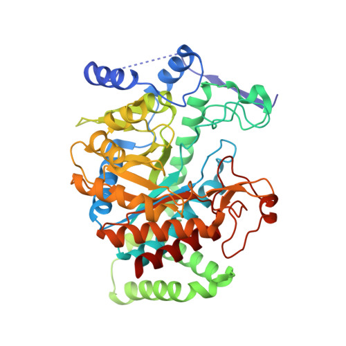
    Allosteric regulation of protein function is critical for metabolic control. Binding of allosteric effectors elicits a functional change in a remote ligand binding site on a protein by altering the equilibrium between different forms in the protein ensemble. 3-Deoxy-d-arabino-heptulosonate 7-phosphate synthase (DAH7PS) catalyzes the first step in the shikimate pathway, which is responsible for the biosynthesis of aromatic amino acids Trp, Phe, and Tyr. Feedback regulation by the aromatic amino acids is important for controlling the cellular levels of the aromatic amino acids, and many organisms have two or more DAH7PS isozymes that show differing sensitivities to aromatic compounds. Mycobacterium tuberculosis expresses a single DAH7PS that is insensitive to the presence of a single amino acid yet shows extraordinary synergistic inhibition by combinations of the pathway end products Trp and Phe. The Trp+Phe-bound structure for M. tuberculosis DAH7PS, showing two separate binding sites occupied by Trp and Phe for each monomer of the tetrameric protein, was obtained by cocrystallization. Comparison of this structure with the ligand-free M. tuberculosis DAH7PS demonstrates that there is no significant change in conformation upon ligand binding, suggesting that contributions from altered dynamic properties of the enzyme may account for the allosteric inhibition. Isothermal titration calorimetry experiments demonstrate that the inhibitor binding sites are in direct communication. Molecular dynamics simulations reveal different changes in dynamic fluctuations upon single ligand binding compared to dual ligand binding. These changes account for the cross-talk between inhibitor binding sites and the active site, simultaneously potentiating both dual ligand binding and diminution of catalytic function.


  • Organizational Affiliation

    Biomolecular Interaction Centre, Department of Chemistry, University of Canterbury, Christchurch, New Zealand.


Macromolecules
Find similar proteins by:  (by identity cutoff)  |  3D Structure
Entity ID: 1
MoleculeChains Sequence LengthOrganismDetailsImage
Probable 3-deoxy-D-arabino-heptulosonate 7-phosphate synthase AroG
A, B
462Mycobacterium tuberculosis H37RvMutation(s): 0 
Gene Names: aroGRv2178c
EC: 2.5.1.54
UniProt
Find proteins for O53512 (Mycobacterium tuberculosis (strain ATCC 25618 / H37Rv))
Explore O53512 
Go to UniProtKB:  O53512
Entity Groups  
Sequence Clusters30% Identity50% Identity70% Identity90% Identity95% Identity100% Identity
UniProt GroupO53512
Sequence Annotations
Expand
  • Reference Sequence
Small Molecules
Ligands 7 Unique
IDChains Name / Formula / InChI Key2D Diagram3D Interactions
TRP
Query on TRP

Download Ideal Coordinates CCD File 
E [auth A],
O [auth B]
TRYPTOPHAN
C11 H12 N2 O2
QIVBCDIJIAJPQS-VIFPVBQESA-N
PHE
Query on PHE

Download Ideal Coordinates CCD File 
D [auth A],
N [auth B]
PHENYLALANINE
C9 H11 N O2
COLNVLDHVKWLRT-QMMMGPOBSA-N
SO4
Query on SO4

Download Ideal Coordinates CCD File 
G [auth A],
Q [auth B],
R [auth B],
S [auth B]
SULFATE ION
O4 S
QAOWNCQODCNURD-UHFFFAOYSA-L
PO4
Query on PO4

Download Ideal Coordinates CCD File 
F [auth A],
P [auth B]
PHOSPHATE ION
O4 P
NBIIXXVUZAFLBC-UHFFFAOYSA-K
GOL
Query on GOL

Download Ideal Coordinates CCD File 
H [auth A]
I [auth A]
K [auth A]
M [auth B]
U [auth B]
H [auth A],
I [auth A],
K [auth A],
M [auth B],
U [auth B],
V [auth B],
W [auth B],
X [auth B]
GLYCEROL
C3 H8 O3
PEDCQBHIVMGVHV-UHFFFAOYSA-N
MN
Query on MN

Download Ideal Coordinates CCD File 
C [auth A],
L [auth B]
MANGANESE (II) ION
Mn
WAEMQWOKJMHJLA-UHFFFAOYSA-N
CL
Query on CL

Download Ideal Coordinates CCD File 
J [auth A],
T [auth B]
CHLORIDE ION
Cl
VEXZGXHMUGYJMC-UHFFFAOYSA-M
Experimental Data & Validation

Experimental Data

  • Method: X-RAY DIFFRACTION
  • Resolution: 1.95 Å
  • R-Value Free: 0.168 
  • R-Value Work: 0.153 
  • R-Value Observed: 0.154 
  • Space Group: P 32 2 1
Unit Cell:
Length ( Å )Angle ( ˚ )
a = 207.56α = 90
b = 207.56β = 90
c = 66.977γ = 120
Software Package:
Software NamePurpose
d*TREKdata scaling
MOLREPphasing
REFMACrefinement
PDB_EXTRACTdata extraction
CrystalCleardata collection
d*TREKdata reduction

Structure Validation

View Full Validation Report


Ligand Structure Quality Assessment 


Entry History 

Deposition Data

Revision History  (Full details and data files)

  • Version 1.0: 2012-01-25
    Type: Initial release
  • Version 1.1: 2013-07-03
    Changes: Database references
  • Version 1.2: 2023-11-01
    Changes: Data collection, Database references, Derived calculations, Refinement description