3RUP

Crystal structure of E.coli biotin carboxylase in complex with two ADP and two Ca ions


Experimental Data Snapshot

  • Method: X-RAY DIFFRACTION
  • Resolution: 1.99 Å
  • R-Value Free: 0.227 
  • R-Value Work: 0.171 
  • R-Value Observed: 0.174 

wwPDB Validation   3D Report Full Report


Ligand Structure Quality Assessment 


This is version 1.4 of the entry. See complete history


Literature

Structural and biochemical studies on the regulation of biotin carboxylase by substrate inhibition and dimerization.

Chou, C.Y.Tong, L.

(2011) J Biol Chem 286: 24417-24425

  • DOI: https://doi.org/10.1074/jbc.M111.220517
  • Primary Citation of Related Structures:  
    3RUP, 3RV3, 3RV4

  • PubMed Abstract: 

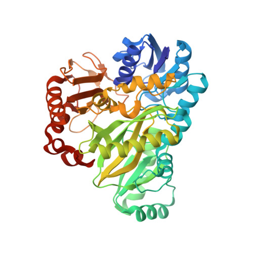
    Biotin carboxylase (BC) activity is shared among biotin-dependent carboxylases and catalyzes the Mg-ATP-dependent carboxylation of biotin using bicarbonate as the CO(2) donor. BC has been studied extensively over the years by structural, kinetic, and mutagenesis analyses. Here we report three new crystal structures of Escherichia coli BC at up to 1.9 Å resolution, complexed with different ligands. Two structures are wild-type BC in complex with two ADP molecules and two Ca(2+) ions or two ADP molecules and one Mg(2+) ion. One ADP molecule is in the position normally taken by the ATP substrate, whereas the other ADP molecule occupies the binding sites of bicarbonate and biotin. One Ca(2+) ion and the Mg(2+) ion are associated with the ADP molecule in the active site, and the other Ca(2+) ion is coordinated by Glu-87, Glu-288, and Asn-290. Our kinetic studies confirm that ATP shows substrate inhibition and that this inhibition is competitive against bicarbonate. The third structure is on the R16E mutant in complex with bicarbonate and Mg-ADP. Arg-16 is located near the dimer interface. The R16E mutant has only a 2-fold loss in catalytic activity compared with the wild-type enzyme. Analytical ultracentrifugation experiments showed that the mutation significantly destabilized the dimer, although the presence of substrates can induce dimer formation. The binding modes of bicarbonate and Mg-ADP are essentially the same as those to the wild-type enzyme. However, the mutation greatly disrupted the dimer interface and caused a large re-organization of the dimer. The structures of these new complexes have implications for the catalysis by BC.


  • Organizational Affiliation

    Department of Life Sciences and Institute of Genome Sciences, National Yang-Ming University, Taipei 112, Taiwan.


Macromolecules
Find similar proteins by:  (by identity cutoff)  |  3D Structure
Entity ID: 1
MoleculeChains Sequence LengthOrganismDetailsImage
Biotin carboxylase
A, B
452Escherichia coli K-12Mutation(s): 0 
Gene Names: accCfabGb3256JW3224
EC: 6.3.4.14 (PDB Primary Data), 6.4.1.2 (PDB Primary Data)
UniProt
Find proteins for P24182 (Escherichia coli (strain K12))
Explore P24182 
Go to UniProtKB:  P24182
Entity Groups  
Sequence Clusters30% Identity50% Identity70% Identity90% Identity95% Identity100% Identity
UniProt GroupP24182
Sequence Annotations
Expand
  • Reference Sequence
Small Molecules
Ligands 3 Unique
IDChains Name / Formula / InChI Key2D Diagram3D Interactions
ADP
Query on ADP

Download Ideal Coordinates CCD File 
C [auth A],
D [auth A],
H [auth B],
I [auth B]
ADENOSINE-5'-DIPHOSPHATE
C10 H15 N5 O10 P2
XTWYTFMLZFPYCI-KQYNXXCUSA-N
CA
Query on CA

Download Ideal Coordinates CCD File 
E [auth A],
F [auth A],
J [auth B],
K [auth B]
CALCIUM ION
Ca
BHPQYMZQTOCNFJ-UHFFFAOYSA-N
CL
Query on CL

Download Ideal Coordinates CCD File 
G [auth A],
L [auth B],
M [auth B]
CHLORIDE ION
Cl
VEXZGXHMUGYJMC-UHFFFAOYSA-M
Binding Affinity Annotations 
IDSourceBinding Affinity
ADP PDBBind:  3RUP Ki: 2.70e+7 (nM) from 1 assay(s)
Experimental Data & Validation

Experimental Data

  • Method: X-RAY DIFFRACTION
  • Resolution: 1.99 Å
  • R-Value Free: 0.227 
  • R-Value Work: 0.171 
  • R-Value Observed: 0.174 
  • Space Group: C 1 2 1
Unit Cell:
Length ( Å )Angle ( ˚ )
a = 170.18α = 90
b = 58.843β = 94.24
c = 85.083γ = 90
Software Package:
Software NamePurpose
SCALEPACKdata scaling
PHASERphasing
REFMACrefinement
PDB_EXTRACTdata extraction
DENZOdata reduction

Structure Validation

View Full Validation Report


Ligand Structure Quality Assessment 


Entry History 

Deposition Data

Revision History  (Full details and data files)

  • Version 1.0: 2011-05-18
    Type: Initial release
  • Version 1.1: 2011-07-13
    Changes: Version format compliance
  • Version 1.2: 2011-07-20
    Changes: Database references
  • Version 1.3: 2017-10-25
    Changes: Author supporting evidence, Refinement description
  • Version 1.4: 2024-02-28
    Changes: Data collection, Database references, Derived calculations, Refinement description