3RUL

New strategy to analyze structures of glycopeptide-target complexes


Experimental Data Snapshot

  • Method: X-RAY DIFFRACTION
  • Resolution: 2.50 Å
  • R-Value Free: 0.277 
  • R-Value Work: 0.243 
  • R-Value Observed: 0.244 

wwPDB Validation   3D Report Full Report


Ligand Structure Quality Assessment 


This is version 2.2 of the entry. See complete history


Literature

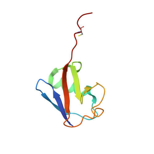
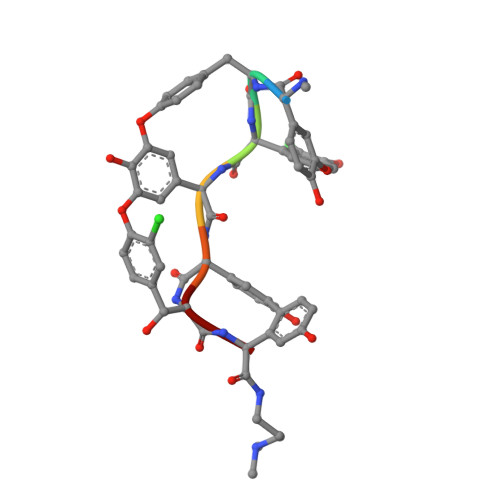
A carrier protein strategy yields the structure of dalbavancin.

Economou, N.J.Nahoum, V.Weeks, S.D.Grasty, K.C.Zentner, I.J.Townsend, T.M.Bhuiya, M.W.Cocklin, S.Loll, P.J.

(2012) J Am Chem Soc 134: 4637-4645

  • DOI: https://doi.org/10.1021/ja208755j
  • Primary Citation of Related Structures:  
    3RUL, 3RUM, 3RUN

  • PubMed Abstract: 

    Many large natural product antibiotics act by specifically binding and sequestering target molecules found on bacterial cells. We have developed a new strategy to expedite the structural analysis of such antibiotic-target complexes, in which we covalently link the target molecules to carrier proteins, and then crystallize the entire carrier-target-antibiotic complex. Using native chemical ligation, we have linked the Lys-D-Ala-D-Ala binding epitope for glycopeptide antibiotics to three different carrier proteins. We show that recognition of this peptide by multiple antibiotics is not compromised by the presence of the carrier protein partner, and use this approach to determine the first-ever crystal structure for the new therapeutic dalbavancin. We also report the first crystal structure of an asymmetric ristocetin antibiotic dimer, as well as the structure of vancomycin bound to a carrier-target fusion. The dalbavancin structure reveals an antibiotic molecule that has closed around its binding partner; it also suggests mechanisms by which the drug can enhance its half-life by binding to serum proteins, and be targeted to bacterial membranes. Notably, the carrier protein approach is not limited to peptide ligands such as Lys-D-Ala-D-Ala, but is applicable to a diverse range of targets. This strategy is likely to yield structural insights that accelerate new therapeutic development.


  • Organizational Affiliation

    Department of Biochemistry and Molecular Biology, Drexel University College of Medicine, Philadelphia, Pennsylvania 19102, United States.


Macromolecules
Find similar proteins by:  (by identity cutoff)  |  3D Structure
Entity ID: 1
MoleculeChains Sequence LengthOrganismDetailsImage
Ubiquitin
A, B, C, D
79Homo sapiensMutation(s): 0 
Gene Names: UBC
UniProt & NIH Common Fund Data Resources
Find proteins for P0CG48 (Homo sapiens)
Explore P0CG48 
Go to UniProtKB:  P0CG48
PHAROS:  P0CG48
GTEx:  ENSG00000150991 
Entity Groups  
Sequence Clusters30% Identity50% Identity70% Identity90% Identity95% Identity100% Identity
UniProt GroupP0CG48
Sequence Annotations
Expand
  • Reference Sequence

Find similar proteins by:  Sequence   |   3D Structure  

Entity ID: 2
MoleculeChains Sequence LengthOrganismDetailsImage
Dalbavancin
E, F, G, H
7N/AMutation(s): 0 
Sequence Annotations
Expand
  • Reference Sequence
Small Molecules
Ligands 5 Unique
IDChains Name / Formula / InChI Key2D Diagram3D Interactions
M12
Query on M12

Download Ideal Coordinates CCD File 
P [auth E],
S [auth F],
V [auth G],
Y [auth H]
10-METHYLUNDECANOIC ACID
C12 H24 O2
QJRRBVNPIKYRQJ-UHFFFAOYSA-N
N1L
Query on N1L

Download Ideal Coordinates CCD File 
N [auth E],
Q [auth F],
T [auth G],
W [auth H]
2-amino-2-deoxy-beta-D-glucopyranuronic acid
C6 H11 N O6
CRIPFXSBMSGPKB-LVBGKUAWSA-N
MAN
Query on MAN

Download Ideal Coordinates CCD File 
O [auth E],
R [auth F],
U [auth G],
X [auth H]
alpha-D-mannopyranose
C6 H12 O6
WQZGKKKJIJFFOK-PQMKYFCFSA-N
TLA
Query on TLA

Download Ideal Coordinates CCD File 
I [auth A],
J [auth A],
K [auth B],
L [auth B]
L(+)-TARTARIC ACID
C4 H6 O6
FEWJPZIEWOKRBE-JCYAYHJZSA-N
CL
Query on CL

Download Ideal Coordinates CCD File 
M [auth B]CHLORIDE ION
Cl
VEXZGXHMUGYJMC-UHFFFAOYSA-M
Modified Residues  2 Unique
IDChains TypeFormula2D DiagramParent
CCS
Query on CCS
A, B, C, D
L-PEPTIDE LINKINGC5 H9 N O4 SCYS
OMY
Query on OMY
E, F, G, H
L-PEPTIDE LINKINGC9 H10 Cl N O4TYR
Experimental Data & Validation

Experimental Data

  • Method: X-RAY DIFFRACTION
  • Resolution: 2.50 Å
  • R-Value Free: 0.277 
  • R-Value Work: 0.243 
  • R-Value Observed: 0.244 
  • Space Group: P 21 21 21
Unit Cell:
Length ( Å )Angle ( ˚ )
a = 53.3α = 90
b = 86.25β = 90
c = 107.19γ = 90
Software Package:
Software NamePurpose
XSCALEdata scaling
MOLREPphasing
PHENIXrefinement
PDB_EXTRACTdata extraction
XDSdata reduction

Structure Validation

View Full Validation Report


Ligand Structure Quality Assessment 


Entry History 

Deposition Data

Revision History  (Full details and data files)

  • Version 1.0: 2012-06-06
    Type: Initial release
  • Version 2.0: 2020-07-29
    Type: Remediation
    Reason: Carbohydrate remediation
    Changes: Atomic model, Data collection, Derived calculations, Polymer sequence, Structure summary
  • Version 2.1: 2023-09-13
    Changes: Data collection, Database references, Refinement description, Structure summary
  • Version 2.2: 2023-12-06
    Changes: Data collection, Derived calculations