3RRC

Crystal Structure of Region II from Plasmodium vivax Duffy Binding Protein


Experimental Data Snapshot

  • Method: X-RAY DIFFRACTION
  • Resolution: 1.95 Å
  • R-Value Free: 0.241 
  • R-Value Work: 0.195 
  • R-Value Observed: 0.197 

wwPDB Validation   3D Report Full Report


This is version 1.2 of the entry. See complete history


Literature

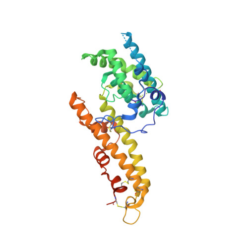
Dimerization of Plasmodium vivax DBP is induced upon receptor binding and drives recognition of DARC.

Batchelor, J.D.Zahm, J.A.Tolia, N.H.

(2011) Nat Struct Mol Biol 18: 908-914

  • DOI: https://doi.org/10.1038/nsmb.2088
  • Primary Citation of Related Structures:  
    3RRC

  • PubMed Abstract: 

    Plasmodium vivax and Plasmodium knowlesi invasion depends on the parasite Duffy-binding protein DBL domain (RII-PvDBP or RII-PkDBP) engaging the Duffy antigen receptor for chemokines (DARC) on red blood cells. Inhibition of this key interaction provides an excellent opportunity for parasite control. There are competing models for whether Plasmodium ligands engage receptors as monomers or dimers, a question whose resolution has profound implications for parasite biology and control. We report crystallographic, solution and functional studies of RII-PvDBP showing that dimerization is required for and driven by receptor engagement. This work provides a unifying framework for prior studies and accounts for the action of naturally acquired blocking antibodies and the mechanism of immune evasion. We show that dimerization is conserved in DBL-domain receptor engagement and propose that receptor-mediated ligand dimerization drives receptor affinity and specificity. Because dimerization is prevalent in signaling, our studies raise the possibility that induced dimerization may activate pathways for invasion.


  • Organizational Affiliation

    Department of Molecular Microbiology, Washington University School of Medicine, Saint Louis, Missouri, USA.


Macromolecules
Find similar proteins by:  (by identity cutoff)  |  3D Structure
Entity ID: 1
MoleculeChains Sequence LengthOrganismDetailsImage
Duffy receptor
A, B
317Plasmodium vivax Sal-1Mutation(s): 0 
Gene Names: PVDR
UniProt
Find proteins for P22290 (Plasmodium vivax (strain Salvador I))
Explore P22290 
Go to UniProtKB:  P22290
Entity Groups  
Sequence Clusters30% Identity50% Identity70% Identity90% Identity95% Identity100% Identity
UniProt GroupP22290
Sequence Annotations
Expand
  • Reference Sequence
Small Molecules
Ligands 2 Unique
IDChains Name / Formula / InChI Key2D Diagram3D Interactions
PO4
Query on PO4

Download Ideal Coordinates CCD File 
I [auth A],
J [auth A],
P [auth B],
Q [auth B]
PHOSPHATE ION
O4 P
NBIIXXVUZAFLBC-UHFFFAOYSA-K
EDO
Query on EDO

Download Ideal Coordinates CCD File 
C [auth A]
D [auth A]
E [auth A]
F [auth A]
G [auth A]
C [auth A],
D [auth A],
E [auth A],
F [auth A],
G [auth A],
H [auth A],
K [auth B],
L [auth B],
M [auth B],
N [auth B],
O [auth B]
1,2-ETHANEDIOL
C2 H6 O2
LYCAIKOWRPUZTN-UHFFFAOYSA-N
Experimental Data & Validation

Experimental Data

  • Method: X-RAY DIFFRACTION
  • Resolution: 1.95 Å
  • R-Value Free: 0.241 
  • R-Value Work: 0.195 
  • R-Value Observed: 0.197 
  • Space Group: P 1 21 1
Unit Cell:
Length ( Å )Angle ( ˚ )
a = 59.5α = 90
b = 66.97β = 108.06
c = 91.83γ = 90
Software Package:
Software NamePurpose
Blu-Icedata collection
SHARPphasing
PHENIXrefinement
XDSdata reduction
XDSdata scaling

Structure Validation

View Full Validation Report


Entry History 

Deposition Data

  • Released Date: 2011-07-13 
  • Deposition Author(s): Tolia, N.H.

Revision History  (Full details and data files)

  • Version 1.0: 2011-07-13
    Type: Initial release
  • Version 1.1: 2011-11-30
    Changes: Database references
  • Version 1.2: 2017-11-08
    Changes: Refinement description