3RLP

Co-crystal structure of the HSP90 ATP binding domain in complex with 4-(2,4-dichloro-5-methoxyphenyl)-6-methylpyrimidin-2-amine


Experimental Data Snapshot

  • Method: X-RAY DIFFRACTION
  • Resolution: 1.70 Å
  • R-Value Free: 0.219 
  • R-Value Work: 0.196 
  • R-Value Observed: 0.197 

wwPDB Validation   3D Report Full Report


Ligand Structure Quality Assessment 


This is version 1.3 of the entry. See complete history


Literature

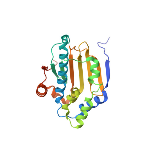
Design strategies to target crystallographic waters applied to the Hsp90 molecular chaperone.

Kung, P.P.Sinnema, P.J.Richardson, P.Hickey, M.J.Gajiwala, K.S.Wang, F.Huang, B.McClellan, G.Wang, J.Maegley, K.Bergqvist, S.Mehta, P.P.Kania, R.

(2011) Bioorg Med Chem Lett 21: 3557-3562

  • DOI: https://doi.org/10.1016/j.bmcl.2011.04.130
  • Primary Citation of Related Structures:  
    3RLP, 3RLQ, 3RLR

  • PubMed Abstract: 

    A series of novel and potent small molecule Hsp90 inhibitors was optimized using X-ray crystal structures. These compounds bind in a deep pocket of the Hsp90 enzyme that is partially comprised by residues Asn51 and Ser52. Displacement of several water molecules observed crystallographically in this pocket using rule-based strategies led to significant improvements in inhibitor potency. An optimized inhibitor (compound 17) exhibited potent Hsp90 inhibition in ITC, biochemical, and cell-based assays (K(d)=1.3 nM, K(i)=15 nM, and cellular IC(50)=0.5 μM).


  • Organizational Affiliation

    Pfizer Worldwide Research and Development, La Jolla Laboratories, San Diego, CA 92121, USA. peipei.kung@pfizer.com


Macromolecules
Find similar proteins by:  (by identity cutoff)  |  3D Structure
Entity ID: 1
MoleculeChains Sequence LengthOrganismDetailsImage
Heat shock protein HSP 90-alpha
A, B
226Homo sapiensMutation(s): 0 
Gene Names: HSP90AA1HSP90AHSPC1HSPCA
UniProt & NIH Common Fund Data Resources
Find proteins for P07900 (Homo sapiens)
Explore P07900 
Go to UniProtKB:  P07900
PHAROS:  P07900
GTEx:  ENSG00000080824 
Entity Groups  
Sequence Clusters30% Identity50% Identity70% Identity90% Identity95% Identity100% Identity
UniProt GroupP07900
Sequence Annotations
Expand
  • Reference Sequence
Small Molecules
Binding Affinity Annotations 
IDSourceBinding Affinity
3RP Binding MOAD:  3RLP Kd: 84 (nM) from 1 assay(s)
PDBBind:  3RLP Kd: 84 (nM) from 1 assay(s)
BindingDB:  3RLP IC50: 1.00e+4 (nM) from 1 assay(s)
Experimental Data & Validation

Experimental Data

  • Method: X-RAY DIFFRACTION
  • Resolution: 1.70 Å
  • R-Value Free: 0.219 
  • R-Value Work: 0.196 
  • R-Value Observed: 0.197 
  • Space Group: P 21 21 21
Unit Cell:
Length ( Å )Angle ( ˚ )
a = 45.583α = 90
b = 79.305β = 90
c = 118.471γ = 90
Software Package:
Software NamePurpose
ADSCdata collection
CNXrefinement
HKL-2000data reduction
SCALEPACKdata scaling
CNXphasing

Structure Validation

View Full Validation Report


Ligand Structure Quality Assessment 


Entry History 

Revision History  (Full details and data files)

  • Version 1.0: 2011-06-08
    Type: Initial release
  • Version 1.1: 2011-07-13
    Changes: Version format compliance
  • Version 1.2: 2015-02-04
    Changes: Structure summary
  • Version 1.3: 2024-02-28
    Changes: Data collection, Database references, Derived calculations