3RLG

Crystal structure of Loxosceles intermedia phospholipase D isoform 1 H12A mutant


Experimental Data Snapshot

  • Method: X-RAY DIFFRACTION
  • Resolution: 1.60 Å
  • R-Value Free: 0.198 
  • R-Value Work: 0.164 
  • R-Value Observed: 0.166 

wwPDB Validation   3D Report Full Report


This is version 1.1 of the entry. See complete history


Literature

Crystallization and preliminary X-ray diffraction analysis of a class II phospholipase D from Loxosceles intermedia venom.

Ullah, A.de Giuseppe, P.O.Murakami, M.T.Trevisan-Silva, D.Wille, A.C.Chaves-Moreira, D.Gremski, L.H.da Silveira, R.B.Sennf-Ribeiro, A.Chaim, O.M.Veiga, S.S.Arni, R.K.

(2011) Acta Crystallogr Sect F Struct Biol Cryst Commun 67: 234-236

  • DOI: https://doi.org/10.1107/S1744309110050931
  • Primary Citation of Related Structures:  
    3RLG

  • PubMed Abstract: 

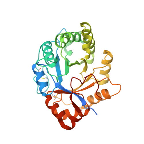
    Phospholipases D are the major dermonecrotic component of Loxosceles venom and catalyze the hydrolysis of phospholipids, resulting in the formation of lipid mediators such as ceramide-1-phosphate and lysophosphatidic acid which can induce pathological and biological responses. Phospholipases D can be classified into two classes depending on their catalytic efficiency and the presence of an additional disulfide bridge. In this work, both wild-type and H12A-mutant forms of the class II phospholipase D from L. intermedia venom were crystallized. Wild-type and H12A-mutant crystals were grown under very similar conditions using PEG 200 as a precipitant and belonged to space group P12(1)1, with unit-cell parameters a = 50.1, b = 49.5, c = 56.5 Å, β = 105.9°. Wild-type and H12A-mutant crystals diffracted to maximum resolutions of 1.95 and 1.60 Å, respectively.


  • Organizational Affiliation

    Centro Multiusuário de Inovação Biomolecular, Departamento de Física, Universidade Estadual Paulista (UNESP), São José do Rio Preto-SP, Brazil.


Macromolecules
Find similar proteins by:  (by identity cutoff)  |  3D Structure
Entity ID: 1
MoleculeChains Sequence LengthOrganismDetailsImage
Sphingomyelin phosphodiesterase D LiSicTox-alphaIA1a302Loxosceles intermediaMutation(s): 1 
EC: 3.1.4.41
UniProt
Find proteins for P0CE80 (Loxosceles intermedia)
Explore P0CE80 
Go to UniProtKB:  P0CE80
Entity Groups  
Sequence Clusters30% Identity50% Identity70% Identity90% Identity95% Identity100% Identity
UniProt GroupP0CE80
Sequence Annotations
Expand
  • Reference Sequence
Small Molecules
Ligands 4 Unique
IDChains Name / Formula / InChI Key2D Diagram3D Interactions
PGE
Query on PGE

Download Ideal Coordinates CCD File 
E [auth A],
H [auth A]
TRIETHYLENE GLYCOL
C6 H14 O4
ZIBGPFATKBEMQZ-UHFFFAOYSA-N
PEG
Query on PEG

Download Ideal Coordinates CCD File 
D [auth A],
I [auth A]
DI(HYDROXYETHYL)ETHER
C4 H10 O3
MTHSVFCYNBDYFN-UHFFFAOYSA-N
EDO
Query on EDO

Download Ideal Coordinates CCD File 
C [auth A],
F [auth A],
G [auth A],
J [auth A]
1,2-ETHANEDIOL
C2 H6 O2
LYCAIKOWRPUZTN-UHFFFAOYSA-N
MG
Query on MG

Download Ideal Coordinates CCD File 
B [auth A]MAGNESIUM ION
Mg
JLVVSXFLKOJNIY-UHFFFAOYSA-N
Experimental Data & Validation

Experimental Data

  • Method: X-RAY DIFFRACTION
  • Resolution: 1.60 Å
  • R-Value Free: 0.198 
  • R-Value Work: 0.164 
  • R-Value Observed: 0.166 
  • Space Group: P 1 21 1
Unit Cell:
Length ( Å )Angle ( ˚ )
a = 49.575α = 90
b = 49.455β = 105.56
c = 56.395γ = 90
Software Package:
Software NamePurpose
DENZOdata reduction
SCALEPACKdata scaling
MOLREPphasing
REFMACrefinement
PDB_EXTRACTdata extraction
MAR345dtbdata collection

Structure Validation

View Full Validation Report


Entry History 

Deposition Data

Revision History  (Full details and data files)

  • Version 1.0: 2011-08-24
    Type: Initial release
  • Version 1.1: 2023-09-13
    Changes: Data collection, Database references, Derived calculations, Refinement description