3RL8

Crystal structure of hDLG1-PDZ2 complexed with APC


Experimental Data Snapshot

  • Method: X-RAY DIFFRACTION
  • Resolution: 2.20 Å
  • R-Value Free: 0.245 
  • R-Value Work: 0.204 
  • R-Value Observed: 0.206 

wwPDB Validation   3D Report Full Report


This is version 1.2 of the entry. See complete history


Literature

Molecular basis for the recognition of adenomatous polyposis coli by the Discs Large 1 protein.

Zhang, Z.Li, H.Chen, L.Lu, X.Zhang, J.Xu, P.Lin, K.Wu, G.

(2011) PLoS One 6: e23507-e23507

  • DOI: https://doi.org/10.1371/journal.pone.0023507
  • Primary Citation of Related Structures:  
    3RL7, 3RL8

  • PubMed Abstract: 

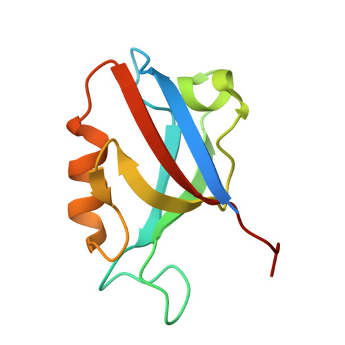
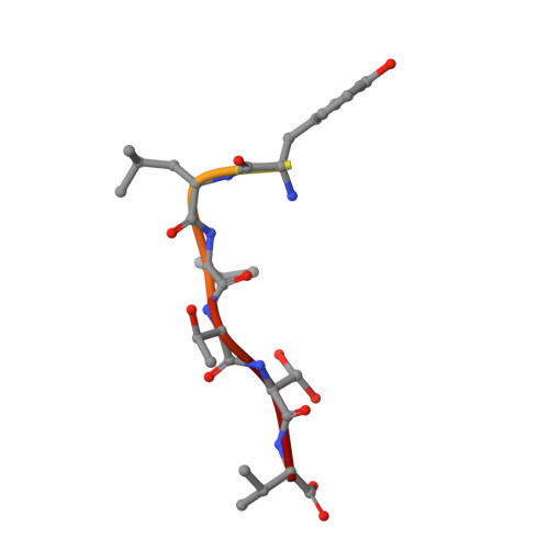
    The human Discs Large 1 (DLG1) protein uses two of its three PDZ domains to interact with the C-terminal peptide of the Adenomatous Polyposis Coli (APC) tumor suppressor protein. The DLG1/APC complex inhibits the cell cycle progression from the G0/G1 to the S phase, regulates epithelial cell migration and morphogenesis, and is required for polarization of the microtubule cytoskeleton. However, the molecular details of how DLG1 recognizes APC is not clear. In this study, we performed biochemical and biophysical assays to investigate the interactions between PDZ domains of DLG1 and the C-terminal peptide of APC. In addition, we determined the crystal structures of the PDZ1 and PDZ2 domains of DLG1 each in complex with the C-terminal 11-residue peptide of APC. Our biochemical, biophysical, and structural results revealed structural elements and residues on PDZ1 and PDZ2 domains of DLG1 and on APC crucial for their mutual interaction. In particular, our results show that the β2/β3 loops of PDZ1 and PDZ2 play important roles in contributing to the binding affinities between PDZ domains and APC, through interacting with the residues upstream of the canonical PDZ-binding S/T-X-V motif. The results provide new insights into the binding mode of a defined C-terminal segment of APC by the PDZ domains of DLG1.


  • Organizational Affiliation

    State Key Laboratory of Microbial Metabolism, and School of Life Sciences and Biotechnology, Shanghai Jiao Tong University, Shanghai, China.


Macromolecules
Find similar proteins by:  (by identity cutoff)  |  3D Structure
Entity ID: 1
MoleculeChains Sequence LengthOrganismDetailsImage
Disks large homolog 1
A, B, C, D, E
105Homo sapiensMutation(s): 0 
Gene Names: DLG1
UniProt & NIH Common Fund Data Resources
Find proteins for Q12959 (Homo sapiens)
Explore Q12959 
Go to UniProtKB:  Q12959
PHAROS:  Q12959
GTEx:  ENSG00000075711 
Entity Groups  
Sequence Clusters30% Identity50% Identity70% Identity90% Identity95% Identity100% Identity
UniProt GroupQ12959
Sequence Annotations
Expand
  • Reference Sequence

Find similar proteins by:  Sequence   |   3D Structure  

Entity ID: 2
MoleculeChains Sequence LengthOrganismDetailsImage
11-mer peptide from Adenomatous polyposis coli protein11Homo sapiensMutation(s): 0 
UniProt & NIH Common Fund Data Resources
Find proteins for P25054 (Homo sapiens)
Explore P25054 
Go to UniProtKB:  P25054
PHAROS:  P25054
GTEx:  ENSG00000134982 
Entity Groups  
Sequence Clusters30% Identity50% Identity70% Identity90% Identity95% Identity100% Identity
UniProt GroupP25054
Sequence Annotations
Expand
  • Reference Sequence
Experimental Data & Validation

Experimental Data

  • Method: X-RAY DIFFRACTION
  • Resolution: 2.20 Å
  • R-Value Free: 0.245 
  • R-Value Work: 0.204 
  • R-Value Observed: 0.206 
  • Space Group: P 1 21 1
Unit Cell:
Length ( Å )Angle ( ˚ )
a = 67.806α = 90
b = 52.502β = 102.42
c = 87.426γ = 90
Software Package:
Software NamePurpose
SCALEPACKdata scaling
PHASERphasing
REFMACrefinement
PDB_EXTRACTdata extraction

Structure Validation

View Full Validation Report


Entry History 

Deposition Data

Revision History  (Full details and data files)

  • Version 1.0: 2011-12-14
    Type: Initial release
  • Version 1.1: 2016-12-21
    Changes: Structure summary
  • Version 1.2: 2023-11-01
    Changes: Data collection, Database references, Refinement description