3RIU

Crystal structure of Drosophila hexameric C3PO formed by truncated Translin and Trax


Experimental Data Snapshot

  • Method: X-RAY DIFFRACTION
  • Resolution: 3.40 Å
  • R-Value Free: 0.338 
  • R-Value Work: 0.271 
  • R-Value Observed: 0.274 

wwPDB Validation   3D Report Full Report


This is version 1.2 of the entry. See complete history


Literature

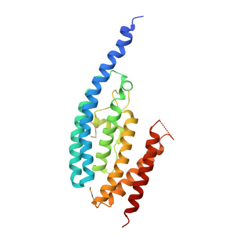
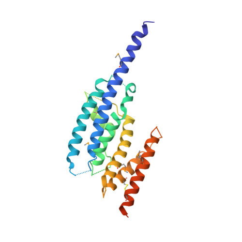
Multimeric assembly and biochemical characterization of the Trax-translin endonuclease complex.

Tian, Y.Simanshu, D.K.Ascano, M.Diaz-Avalos, R.Park, A.Y.Juranek, S.A.Rice, W.J.Yin, Q.Robinson, C.V.Tuschl, T.Patel, D.J.

(2011) Nat Struct Mol Biol 18: 658-664

  • DOI: https://doi.org/10.1038/nsmb.2069
  • Primary Citation of Related Structures:  
    3RIU

  • PubMed Abstract: 

    Trax-translin heteromers, also known as C3PO, have been proposed to activate the RNA-induced silencing complex (RISC) by facilitating endonucleolytic cleavage of the siRNA passenger strand. We report on the crystal structure of hexameric Drosophila C3PO formed by truncated translin and Trax, along with electron microscopic and mass spectrometric studies on octameric C3PO formed by full-length translin and Trax. Our studies establish that Trax adopts the translin fold, possesses catalytic centers essential for C3PO's endoRNase activity and interacts extensively with translin to form an octameric assembly. The catalytic pockets of Trax subunits are located within the interior chamber of the octameric scaffold. Truncated C3PO, like full-length C3PO, shows endoRNase activity that leaves 3'-hydroxyl-cleaved ends. We have measured the catalytic activity of C3PO and shown it to cleave almost stoichiometric amounts of substrate per second.


  • Organizational Affiliation

    Structural Biology Program, Memorial Sloan-Kettering Cancer Center, New York, New York, USA. Graduate Program in Neuroscience, Weill Medical College of Cornell University, New York, New York, USA.


Macromolecules
Find similar proteins by:  (by identity cutoff)  |  3D Structure
Entity ID: 1
MoleculeChains Sequence LengthOrganismDetailsImage
Translin
A, B
218Drosophila melanogasterMutation(s): 0 
Gene Names: CG11761Dmel_CG11761translintrsn
UniProt
Find proteins for Q7JVK6 (Drosophila melanogaster)
Explore Q7JVK6 
Go to UniProtKB:  Q7JVK6
Entity Groups  
Sequence Clusters30% Identity50% Identity70% Identity90% Identity95% Identity100% Identity
UniProt GroupQ7JVK6
Sequence Annotations
Expand
  • Reference Sequence
Find similar proteins by:  (by identity cutoff)  |  3D Structure
Entity ID: 2
MoleculeChains Sequence LengthOrganismDetailsImage
Translin associated factor X, isoform B269Drosophila melanogasterMutation(s): 0 
Gene Names: Dmel_CG5063Trax
UniProt
Find proteins for Q9VF77 (Drosophila melanogaster)
Explore Q9VF77 
Go to UniProtKB:  Q9VF77
Entity Groups  
Sequence Clusters30% Identity50% Identity70% Identity90% Identity95% Identity100% Identity
UniProt GroupQ9VF77
Sequence Annotations
Expand
  • Reference Sequence
Small Molecules
Modified Residues  1 Unique
IDChains TypeFormula2D DiagramParent
MSE
Query on MSE
A, B
L-PEPTIDE LINKINGC5 H11 N O2 SeMET
Experimental Data & Validation

Experimental Data

  • Method: X-RAY DIFFRACTION
  • Resolution: 3.40 Å
  • R-Value Free: 0.338 
  • R-Value Work: 0.271 
  • R-Value Observed: 0.274 
  • Space Group: P 61 2 2
Unit Cell:
Length ( Å )Angle ( ˚ )
a = 196.03α = 90
b = 196.03β = 90
c = 155.189γ = 120
Software Package:
Software NamePurpose
DENZOdata reduction
SCALEPACKdata scaling
SHARPphasing
PHENIXrefinement
PDB_EXTRACTdata extraction

Structure Validation

View Full Validation Report


Entry History 

Deposition Data

Revision History  (Full details and data files)

  • Version 1.0: 2011-05-11
    Type: Initial release
  • Version 1.1: 2011-07-13
    Changes: Version format compliance
  • Version 1.2: 2017-11-08
    Changes: Refinement description