3RIP

Crystal Structure of human gamma-tubulin complex protein 4 (GCP4)


Experimental Data Snapshot

  • Method: X-RAY DIFFRACTION
  • Resolution: 2.30 Å
  • R-Value Free: 0.260 
  • R-Value Work: 0.227 
  • R-Value Observed: 0.228 

wwPDB Validation   3D Report Full Report


This is version 1.3 of the entry. See complete history


Literature

Crystal structure of gamma-tubulin complex protein GCP4 provides insight into microtubule nucleation.

Guillet, V.Knibiehler, M.Gregory-Pauron, L.Remy, M.H.Chemin, C.Raynaud-Messina, B.Bon, C.Kollman, J.M.Agard, D.A.Merdes, A.Mourey, L.

(2011) Nat Struct Mol Biol 18: 915-919

  • DOI: https://doi.org/10.1038/nsmb.2083
  • Primary Citation of Related Structures:  
    3RIP

  • PubMed Abstract: 

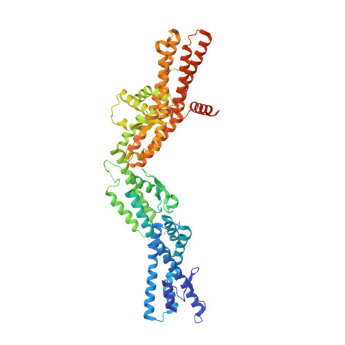
    Microtubule nucleation in all eukaryotes involves γ-tubulin small complexes (γTuSCs) that comprise two molecules of γ-tubulin bound to γ-tubulin complex proteins (GCPs) GCP2 and GCP3. In many eukaryotes, multiple γTuSCs associate with GCP4, GCP5 and GCP6 into large γ-tubulin ring complexes (γTuRCs). Recent cryo-EM studies indicate that a scaffold similar to γTuRCs is formed by lateral association of γTuSCs, with the C-terminal regions of GCP2 and GCP3 binding γ-tubulin molecules. However, the exact role of GCPs in microtubule nucleation remains unknown. Here we report the crystal structure of human GCP4 and show that its C-terminal domain binds directly to γ-tubulin. The human GCP4 structure is the prototype for all GCPs, as it can be precisely positioned within the γTuSC envelope, revealing the nature of protein-protein interactions and conformational changes regulating nucleation activity.


  • Organizational Affiliation

    Institut de Pharmacologie et de Biologie Structurale, Centre National de la Recherche Scientifique, Toulouse, France.


Macromolecules
Find similar proteins by:  (by identity cutoff)  |  3D Structure
Entity ID: 1
MoleculeChains Sequence LengthOrganismDetailsImage
Gamma-tubulin complex component 4677Homo sapiensMutation(s): 0 
Gene Names: TUBGCP476PGCP4
UniProt & NIH Common Fund Data Resources
Find proteins for Q9UGJ1 (Homo sapiens)
Explore Q9UGJ1 
Go to UniProtKB:  Q9UGJ1
PHAROS:  Q9UGJ1
GTEx:  ENSG00000137822 
Entity Groups  
Sequence Clusters30% Identity50% Identity70% Identity90% Identity95% Identity100% Identity
UniProt GroupQ9UGJ1
Sequence Annotations
Expand
  • Reference Sequence
Small Molecules
Ligands 2 Unique
IDChains Name / Formula / InChI Key2D Diagram3D Interactions
MRD
Query on MRD

Download Ideal Coordinates CCD File 
B [auth A](4R)-2-METHYLPENTANE-2,4-DIOL
C6 H14 O2
SVTBMSDMJJWYQN-RXMQYKEDSA-N
GOL
Query on GOL

Download Ideal Coordinates CCD File 
C [auth A]
D [auth A]
E [auth A]
F [auth A]
G [auth A]
C [auth A],
D [auth A],
E [auth A],
F [auth A],
G [auth A],
H [auth A],
I [auth A],
J [auth A]
GLYCEROL
C3 H8 O3
PEDCQBHIVMGVHV-UHFFFAOYSA-N
Experimental Data & Validation

Experimental Data

  • Method: X-RAY DIFFRACTION
  • Resolution: 2.30 Å
  • R-Value Free: 0.260 
  • R-Value Work: 0.227 
  • R-Value Observed: 0.228 
  • Space Group: P 63 2 2
Unit Cell:
Length ( Å )Angle ( ˚ )
a = 214.95α = 90
b = 214.95β = 90
c = 128.66γ = 120
Software Package:
Software NamePurpose
ADSCdata collection
PHASERphasing
REFMACrefinement
XDSdata reduction
XSCALEdata scaling

Structure Validation

View Full Validation Report


Entry History 

Deposition Data

Revision History  (Full details and data files)

  • Version 1.0: 2011-07-06
    Type: Initial release
  • Version 1.1: 2011-07-13
    Changes: Version format compliance
  • Version 1.2: 2013-07-03
    Changes: Database references
  • Version 1.3: 2024-04-03
    Changes: Data collection, Database references, Derived calculations, Refinement description